
Author: The Wistar Institute

Paul M. Lieberman, Ph.D.

  • Hilary Koprowski, M.D., Endowed Professor and Program Leader, Gene Expression and Regulation Program, Ellen and Ronald Caplan Cancer Center

  • Director, Center for Chemical Biology & Translational Medicine

Lieberman studies how certain viruses establish a long-term latent infection that can lead to cancer or autoimmune disorders.

Lieberman joined The Wistar Institute in 1995 as an assistant professor. He earned his bachelor’s degree in chemistry from Cornell University and a doctorate in pharmacology/virology from The Johns Hopkins University School of Medicine, which was followed by a postdoctoral fellowship at the University of California, Los Angeles.

Lieberman Chairs the Program in Gene Expression and Regulation at the Ellen and Ronald Caplan Cancer Center at The Wistar Institute. In 2010, Lieberman became the first director of The Wistar Institute Center for Chemical Biology and Translational Medicine. Using the advanced screening technologies of Wistar’s Molecular Screening Facility, the Center enables scientists to identify and characterize new molecules and compounds that hold the most promise for developing into therapeutic drugs for cancer and other diseases.

View Publications

The Lieberman Laboratory

215-898-9491

lieberman@wistar.org

The Lieberman Laboratory

Research in the Lieberman laboratory centers on understanding how the cancer-associated viruses persist in a latent state and increase the risk of cancer and autoimmune disorders. EBV and KSHV establish latent infections that are associated with several human malignancies, including Burkitt’s lymphoma, nasopharyngeal carcinoma, Hodgkin’s disease, and post-transplant lymphoproliferative disorder for EBV, and Kaposi’s Sarcoma for KSHV. EBV has also been implicated in multiple sclerosis and other autoimmune disorders.

Lieberman and his team found that oncogenic viruses can interact with cellular proteins that regulate telomeres—the repetitive DNA sequences found at the ends of chromosomes. Telomeres protect chromosomes from loss of genetic information, and a similar process is thought to preserve the virus during latency. The Lieberman laboratory worked out several biochemical pathways that control the stability, replication, and gene expression patterns of the latent virus. New research also focuses on the epigenetic controls of latent viruses and human telomeres, and how interactions between viruses and telomeres may induce a malignant transformation of the infected cell.

Staff
  • Senior Staff Scientists

    Troy Messick, Ph.D.
    Samantha Soldan, Ph.D.

  • Associate Staff Scientists

    Amit Gurav, Ph.D.
    Urvi Zankharia, Ph.D.

  • Postdoctoral Fellows

    Leonardo Josue Castro-Munoz, Ph.D.
    Chris Chen, Ph.D.
    Chenhe Su, Ph.D.

  • Research Assistants

    Asim Ashgar
    Andreas Wiedmer
    Olga Vladimirova

Research

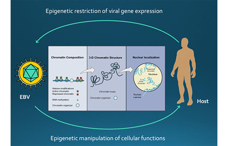
EPIGENETIC CONTROL OF VIRAL LATENCY

Research in the Lieberman laboratory centers on understanding how the cancer-associated viruses, like Epstein-Barr virus (EBV) and Kaposi’s Sarcoma Associated Herpesvirus (KSHV), persist in a latent state and increases the risk of cancer cell evolution. EBV and KSHV establish latent infections that are associated with several human malignancies, including Burkitt’s lymphoma, nasopharyngeal carcinoma, Hodgkin’s disease, and post-transplant lymphoproliferative disorder for EBV, and Kaposi’s Sarcoma for KSHV.

The researchers have recently found that viral DNA replication and maintenance is regulated by interactions with cellular telomere binding proteins. Telomeres are the repetitive DNA sequences found at the ends of chromosomes. Telomeres protect chromosomes from loss of genetic information, and a similar process is thought to preserve the virus during latency. The Lieberman research team has worked out several biochemical pathways that control the stability, replication, and gene expression patterns of the latent virus. They have found that changes in viral chromatin structure alters the cancer-risk associated with latent infection.

VIRUS MODULATION OF HOST CHROMOSOMES

The Lieberman lab continues to study EBV and KSHV genome maintenance proteins, EBNA1 and LANA, respectively. These proteins bind to the viral OriP, but they also bind to the cellular chromosome at unknown sites. The Lieberman lab has identified the cellular chromosome binding sites for both EBNA1 and LANA in latently infected B-lymphocytes. LANA was found to bind to host genes involved in gamma-interferon signaling and LANA may antagonize STAT1/STAT3 binding to host genes important for MHC peptide presentation and processing. EBNA1 may promote higher order structures, including interchromosome linkages that may promote translocations similar to those observed in Burkitt’s lymphoma.

CHROMOSOME CONFORMATION CONTROL OF VIRAL LATENCY

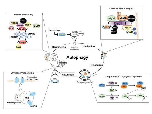
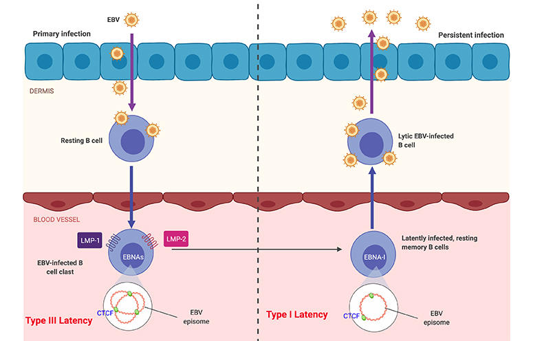
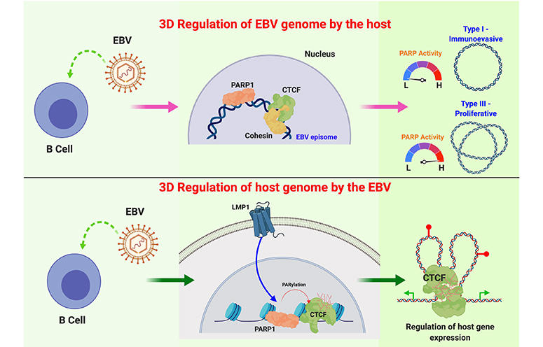
The role of chromosome architecture and higher-ordered structure is also important for genome maintenance. The Lieberman lab has studied the role of chromatin architecture proteins CTCF and cohesins in regulating viral genome structure and gene expression during latent infection. They have shown that CTCF and cohesins mediate long-distance interactions that are important for control of gene expression and maintenance of a stable latent infection. Loss of genome architecture leads to a change in gene expression and a transition from a circular to linear viral genome.

TELOMERE MAINTENANCE AND DYSFUNCTION IN CANCER

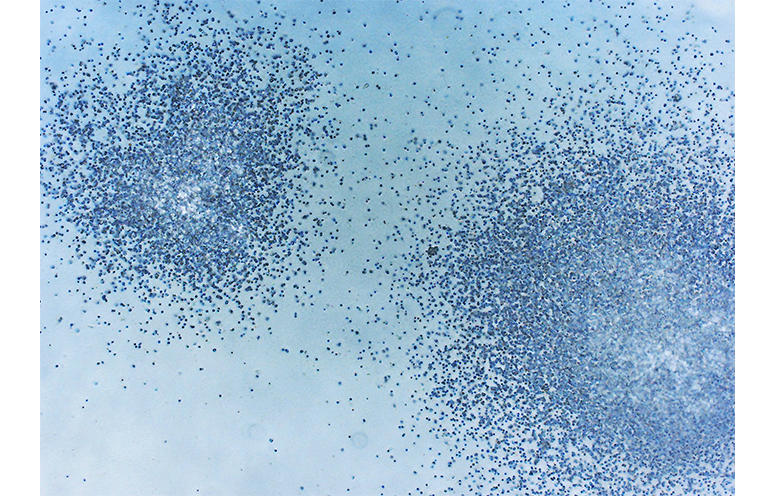
Maintenance of telomere structures that maintain the ends of linear chromosomes is also important for human genome stability. The Lieberman lab is studying the chromatin structure of telomeres and the expression of a telomere repeat-containing non-coding RNA, termed TERRA. They have shown that TERRA is overexpressed in highly proliferating cells in human and mouse cancers. The TERRA form nuclear aggregates in cancer cells in mouse models of medulloblastoma, and TERRA RNA levels were highly over-expressed in human ovarian cancer biopsies. The regulation and function of TERRA expression, and its role in regulating telomere length and stability are the focus of future research.

DRUG DISCOVERY RESEARCH

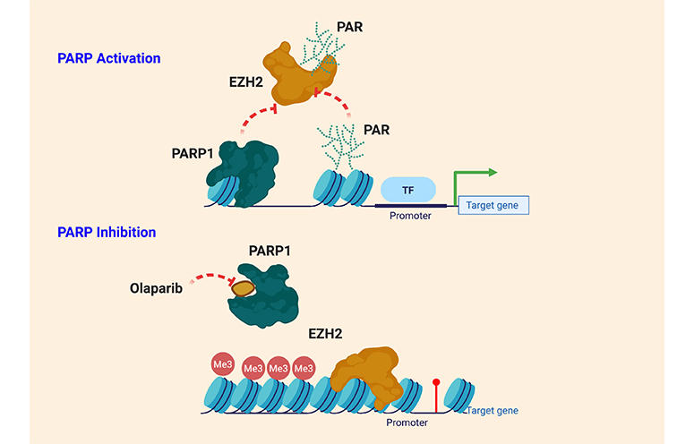
The Lieberman laboratory is also pursuing the development of small molecule inhibitors of the EBV encoded origin binding protein EBNA1. The laboratory is collaborating with structural biologists and medicinal chemists to advance hits into lead compounds for testing in animal models of EBV lymphomagenesis. These small molecules will be considered for further development as inhibitors of EBV-associated malignancies.

Lieberman Lab in the News

Selected Publications

  • Cryo-EM Structure and Functional Studies of EBNA1 Binding to the Family of Repeats and Dyad Symmetry Elements of Epstein-Barr Virus oriP.

    Mei, Y., Messick, T.E., Dheekollu, J., Kim, H.J., Molugu, S., Castro Muñoz, L.J.C., Moiskeenkova-Bell, V., Murakami, K., Lieberman, P.M. “Cryo-EM Structure and Functional Studies of EBNA1 Binding to the Family of Repeats and Dyad Symmetry Elements of Epstein-Barr Virus oriP.” J Virol. 2022 Sep 14;96(17):e0094922. doi: 10.1128/jvi.00949-22. Epub 2022 Aug 29.

  • DAXX-ATRX Regulation of p53 Chromatin Binding and DNA Damage Response.

    Gulve, N., Su, C., Deng, Z., Soldan, S.S., Vladimirova, O., Wickramasinghe, J., Zheng, H., Kossenkov, A.V. , Lieberman, P.M. “DAXXATRX Regulation of p53 Chromatin Binding and DNA Damage Response.” Nat Commun. 2022 Aug 26;13(1):5033. doi: 10.1038/s41467-022-32680-8.

  • Epigenetic Plasticity Enables CNS-Trafficking of EBV-infected B Lymphocytes.

    Soldan, S.S., Su, C., Lamontagne, R.J., Grams, N., Lu, F., Zhang, Y., Gesualdi, J.D., Frase, D.M., Tolvinski, L.E, Martinm, K., et al. “Epigenetic Plasticity Enables CNS-Trafficking of EBV-infected B Lymphocytes.” PLoS Pathog. 2021 Jun 9;17(6):e1009618. doi: 10.1371/journal.ppat.1009618. eCollection 2021 Jun.

  • A Multi-omics Approach to Epstein-Barr Virus Immortalization of B-cells Reveals EBNA1 Chromatin Pioneering Activities Targeting Nucleotide Metabolism.

    Lamontagne, R.J., Samantha, S.S., Su, C., Wiedmer, A., Won, K.J., Lu, F., Goldman, A.R., Wickramasinghe, J., Tang, H.Y., Speicher, D.W., et al. “A Multi-omics Approach to Epstein-Barr Virus Immortalization of B-cells Reveals EBNA1 Chromatin Pioneering Activities Targeting Nucleotide Metabolism.” PLoS Pathog. 2021 Jan 26;17(1):e1009208. doi: 10.1371/journal.ppat.1009208. eCollection 2021 Jan.

  • Cell-cycle-dependent EBNA1-DNA Crosslinking Promotes Replication Termination at oriP and Viral Episome Maintenance.

    Dheekollu, J., Wiedmer, A., Ayyanathan, K., Deakyne, J.S., Messick, T.E., Lieberman, P.M. “Cell-cycle-dependent EBNA1DNA Crosslinking Promotes Replication Termination at oriP and Viral Episome Maintenance.” Cell. 2021 Feb 4;184(3):643-654.e13. doi: 10.1016/j.cell.2020.12.022. Epub 2021 Jan 21.

View Additional Publications

Continue reading

Chengyu Liang, M.D., Ph.D.

Professor and Program Co-Leader, Molecular and Cellular Oncogenesis Program, Ellen and Ronald Caplan Cancer Center

Liang’s research explores basic mechanisms underlying fundamental cellular processes in inflammation, infection, and cancer, broadly focusing on autophagy, organelle homeostasis, genomic stability, membrane trafficking, and virus-host interaction.

Liang obtained her M.D. degree from Qingdao University School of Medicine, China, and her Ph.D. degree in genetics from State University of New York (SUNY) at Stony Brook. She received postdoctoral training in tumor virology at Harvard Medical School in the Department of Microbiology and Molecular Genetics. She established her laboratory in 2009 in the Department of Molecular Microbiology and Immunology, University of Southern California, Keck School of Medicine in Los Angeles, where she was promoted to tenured associate professor in 2015. Liang joined The Wistar Institute as a professor in 2020.

View Publications

The Liang Laboratory

215-898-3862

cliang@wistar.org

The Liang Laboratory

Autophagy (literally “self-eating”) is the natural, regulated mechanism cells use to digest and remove unwanted components. This process influences diverse aspects of cell homeostasis and constitutes a barrier against malignant transformation.

The Liang lab identified a novel autophagy pathway controlled by the tumor suppressor gene UVRAG (UV-radiation Resistance-Associated Gene) that also plays a direct role in DNA repair and chromosomal stability. The group studies autophagy and related pathways in leukemia, colorectal cancer, melanoma pathogenesis and therapy resistance, and in viral persistency.

Staff
  • Postdoctoral Fellows

    Jinghui Liang, Ph.D. 
    Behzad Mansoori, Ph.D.
    Christian Pangilinan, Ph.D
    Qing Zhu, Ph.D.

  • Research Assistants

    Nivedita Rathaur
    Solmaz Shirjang

  • Bioinformatics Technicians

    Robert McElroy
    Jonathan Richards

  • Predoctoral Trainees

    Dongliang Shen
    Lu Zhang

  • Lab Manager

    Dali Nemecio 


Alumni

Liang Jin, Ph.D. (2010) Senior Scientist, China

Zhen Zhao, Ph.D. (2010 – 2012) Assistant Professor, University of Southern California

Lingling Shi, Ph.D. (2011 – 2012) Associate Professor, Guangdong-Hong Kong-Macau Institute of CNS Regeneration, China

Shanshan He, M.D., Ph.D. (2012 – 2015) Senior Scientist, NanoString Technologies, Inc.

Yongfei Yang (2013 – 2017) Associate Professor, Beijing Institute of Technology

Gym-beom Jang (2016 – 2018) Staff Researcher, National Cancer Center, Seoul, Korea

Shun Li, Ph.D. (2016 – 2018) Professor, Chengdu Medical School, Sichuan University

Hongrui Guo (2016 – 2018) Professor, Sichuan Agriculture University

Ying Song (2017-2020) Professor, Weifang Medical College


Available Positions

Motivated recent Ph.D. graduates from related fields with a record of publications are encouraged to inquire about the position below. Contact cliang@wistar.org.

– Postdoctoral Fellow

Research

  • UVRAG in Autophagy, Inflammation, and Cancer

    Download the image here.

    Macroautophagy/autophagy deregulation has been observed in perpetuated inflammation and the proliferation of tumor cells. However, the mechanisms underlying these changes have yet to be well-identified. UVRAG is one of the key players of autophagy, but its role in vivo remained puzzling. Our recent study, published in Nature Communications (Quach, Song et al), utilized a mouse model with inducible expression of a cancer-derived frameshift (FS) mutation in UVRAG that dominant-negatively inhibits wild-type UVRAG, resulting in impaired stimulus-induced autophagy. The systemically compromised autophagy, particularly mitophagy, notably increases inflammation and associated pathologies. Another pivotal result of this study is that it uncovered a “missing molecular link” between age-related decreases in autophagy and aging-induced spontaneous cancers, which could provide new insight into how basal autophagy protects against cancer. Using our mouse model, we observed that suppression of UVRAG accelerated age-related decline in autophagy and impaired autophagic turnover of oncogenic β-catenin, leading to increased proliferation and tumor formation in mice. It remains to be seen whether this age-dependent regulation of β-catenin by autophagy will translate to other autophagy-related cancer models. Nevertheless, our work represents a key advance in understanding how UVRAG and by extension autophagy works in vivo.

  • Autophagy-lysomal Activation in Melanoma Pathogenesis and Drug Resistance

    Download the image here.

    Autophagy, originally described as a lysosome-dependent degradation of cytoplasmic components upon starvation, has since been shown to influence diverse aspects of homeostasis, constituting a barrier against malignant transformation. Despite its inhibitory role in tumor initiation, autophagy is postulated to fuel the growth of established tumors and confers drug resistance, principally as a survival mechanism. In melanoma, where 40–60% of cases have a mutation in BRAF (BRAFV600E), conflicting results have been reported regarding the relationship between autophagy and the mutant, and the interaction between BRAF signaling and autophagy has been ambiguous. Our work, published in 2019 in Nature Communications (Li, et al), demonstrated that in BRAFV600E-melanoma autophagy is induced by BRAF inhibitor (BRAFi), as part of a transcriptional program coordinating lysosome biogenesis/function, mediated by the TFEB transcription factor. TFEB is phosphorylated and thus inactivated by BRAFV600E via its downstream ERK independently of mTORC1. BRAFi disrupt TFEB phosphorylation, allowing its nuclear translocation, which is synergized by increased phosphorylation/inactivation of the ZKSCAN3 transcriptional repressor by JNK2/p38-MAPK. Blockade of BRAFi-induced transcriptional activation of autophagy-lysosomal function in melanoma xenografts causes enhanced tumor progression, EMT-transdifferentiation, metastatic dissemination, and chemoresistance, which is associated with elevated TGF-β levels and enhanced TGF-β signaling. Inhibition of TGF-β signaling restores tumor differentiation and drug responsiveness in melanoma cells. Thus, the “BRAF-TFEB-autophagy-lysosome” axis represents an intrinsic regulatory pathway in BRAF-mutant melanoma, coupling BRAF signaling with TGF-β signaling to drive tumor progression and chemoresistance.

  • Molecular Mechanisms of UV-induced Mutagenesis in Skin Melanoma

    Download the image here.

    Malignant melanoma ranks as the number one cause of death from skin cancers. One striking feature of cutaneous melanoma is the highest mutation burden caused by ultraviolet radiation (UVR) in its genome. Such high UV-signature mutation rate provides huge fitness advantage to cancer cells. However, the molecular mechanisms underlying UV-induced mutagenesis in melanoma remain largely unknown, posing major challenges in the development of cancer treatment with long-term durability of responses. While screening for the molecular target(s) that drive UV signature loads in melanoma genomes, we discovered that UV-exposed skin melanoma patients with higher amounts of UV signature in their genomes had reduced levels of UVRAG; but this was not observed for UV-shielded melanoma, suggesting a potential link of UVRAG to photolesion protection. Our work, published in 2016 in Molecular Cell (Yang et al), further demonstrated that UVRAG plays an integral role in UV-induced DNA damage repair. It localizes to photolesions and associates with DDB1 to promote the assembly and activity of the DDB2-DDB1-Cul4A-Roc1 (CRL4DDB2) ubiquitin ligase complex, leading to efficient XPC recruitment and global genomic NER. UVRAG depletion decreased substrate handover to XPC and conferred UV-damage hypersensitivity. These results identify UVRAG as a regulator of CRL4DDB2-mediated NER and suggest that its expression levels may influence melanoma predisposition. Future studies in the laboratory will focus on investigating the mechanisms protecting against UV-induced mutagenesis, and delineate why they fail to work in melanoma, enabling early risk prediction and prognostication, inspiring new strategy to minimize melanoma susceptibility and tumor fitness.

  • Ultraviolet Radiation and Tanning

    Download the image here.

    Skin pigmentation provides first-line protection against UV radiation (UVR) that increases the risk of skin cancers. However, mechanisms underlying this process remain poorly understood. Our work, published in 2018 in PNAS (Yang, Jang et al), identified the autophagic tumor suppressor UVRAG as a bona fide player in melanosome biogenesis by targeting biogenesis of lysosome-related organelles complex 1 (BLOC-1) independently of autophagy. UVRAG maintains the localization and stability of BLOC-1 to facilitate the sorting/delivery of melanogenic cargoes. Reduced levels of UVRAG rendered cells unresponsive to UVR–α-MSH–MITF signaling and defective melanocyte development in vivo. Moreover, UVRAG-mediated melanogenesis and tanning response were impaired in oncogene driven melanoma. This study represents a description of a noncanonical role of autophagy factor in melanogenic remodeling and also provides mechanistic insights into UVRAG in pigmentation disorder and UV-associated cancer. Future studies will continue to investigate the molecular mechanisms controlling melanosome biogenesis/dynamics and their impact on UV protection in skin cancer.

  • Chromosomal Instability and Cancer Progression/Recurrence

    Download the image here.

    Due to the unacceptable toxicity and ineffectiveness of currently cancer chemotherapy and radiation, the search for new molecular targets is in high gear. Chromosomal instability (CIN) is a hallmark of most aggressive cancers. However, efforts to exploit CIN therapeutically have been hampered by a lack of understanding of molecular mechanisms involved in its regulation. Our objective is to characterize the molecular mechanisms that induce CIN and to identify new therapeutic targets for cancer progression by applying our discovery of a novel molecule in CIN regulation. While we initially thought that UVRAG was mainly a promoter of the autophagy pathway, our continued efforts demonstrated that, in addition to its confirmed role in autophagy activation, UVRAG also plays a direct role in preventing cells from accumulating abnormal chromosomes, which would increase the danger of developing oncogenic mutations. As the maintenance of chromosomal integrity is a fundamental biological process to thwart tumor formation, this property of UVRAG also explains why it is frequently mutated in cancers. Our work, published in 2012 in Developmental Cell (Zhao, Oh et al) have discovered two novel mechanisms by which UVRAG mediates nonhomologous end-joining (NHEJ) and centrosome stability in a spatiotemporally distinct manner, independently of autophagy. These findings will help us expand on this knowledge to investigate in depth how UVRAG impacts genomic integrity and cancer development, and determine the molecular crosstalk between the autophagy machinery and the genome surveillance process of the cells.

  • Bcl-2 Protein Family in Autophagy Inhibition, Cancer Progression and Viral Persistency

    Download the image here.

    Bcl-2, originally identified as a universal inhibitor of apoptotic cell death, has since been implicated in suppressing autophagy, the cell’s quality control mechanism. Our study, published in 2011 in Cell Death and Differentiation (Oh et al), demonstrates that the anti-autophagic aspect of Bcl-2 can function as a promoter of oncogenic growth, independently of its role in apoptosis signaling. It is likely that the increase in Bcl-2 often seen in breast and other cancers might render cells error prone by blunting autophagy, while concomitantly keeping damaged cells alive. The outcome of such a ‘double hit’ of Bcl-2 may synergistically promote tumor growth and increase the chance of cancer development and drug resistance. Moreover, our lab has identified the mechanism by which viruses co-opt the autophagic pathway to establish virulence in vivo. Published in 2009 in PLOS Pathogens (E et al), we presented direct in vivo evidence for the key role of the anti-autophagic aspect of the virally encoded Bcl-2s in the chronic infection of oncogenic gamma-herpesviruses, and proposed that cellular autophagy may have a substantial effect on viral persistence and may influence the in vivo fitness of viruses. This discovery expands upon known antiviral activities of the autophagy machinery, and also suggests new approaches for treating some virally induced diseases.

  • Molecular Mechanism of Autophagy and Intracellular Membrane Trafficking

    Download the image here.

    The homeostasis of all eukaryotic cells depends on their “greening” ability to use a lysosomal pathway known as autophagy to degrade and recycle self-components. Our work, published in 2008 and 2013 in Nature Cell Biology (He, et al), have shown for the first time that autophagic and endosomal maturation are of topological similarity and may have evolved to share associated machinery for lysosomal delivery, which has been further confirmed by other laboratories later on. Our seminal findings showed that UVRAG, originally identified as a UV-protecting molecule, is an important coordinator in autophagic and endosomal trafficking through its interaction with the class C Vps tethering complex. Furthermore, UVRAG recognizes PI3P at the ER, where it couples the ER tether to govern Golgi-ER retrograde transport. Intriguingly, when autophagy is induced, UVRAG undergoes a “partnering shift” from the ER complex to the BECN1 autophagy complex, resulting in concomitant inhibition of Golgi-ER transport and activation of ATG9 autophagic trafficking. This work sets up a mechanism of how cells achieve spatiotemporal fidelity of protein transport and organelle homeostasis, providing insights into trafficking-related diseases.

Staff
  • Postdoctoral Fellows

    Jinghui Liang, Ph.D. 
    Behzad Mansoori, Ph.D.
    Christian Pangilinan, Ph.D
    Qing Zhu, Ph.D.

  • Research Assistants

    Nivedita Rathaur
    Solmaz Shirjang

  • Bioinformatics Technicians

    Robert McElroy
    Jonathan Richards

  • Predoctoral Trainees

    Dongliang Shen
    Lu Zhang

  • Lab Manager

    Dali Nemecio 


Alumni

Liang Jin, Ph.D. (2010) Senior Scientist, China

Zhen Zhao, Ph.D. (2010 – 2012) Assistant Professor, University of Southern California

Lingling Shi, Ph.D. (2011 – 2012) Associate Professor, Guangdong-Hong Kong-Macau Institute of CNS Regeneration, China

Shanshan He, M.D., Ph.D. (2012 – 2015) Senior Scientist, NanoString Technologies, Inc.

Yongfei Yang (2013 – 2017) Associate Professor, Beijing Institute of Technology

Gym-beom Jang (2016 – 2018) Staff Researcher, National Cancer Center, Seoul, Korea

Shun Li, Ph.D. (2016 – 2018) Professor, Chengdu Medical School, Sichuan University

Hongrui Guo (2016 – 2018) Professor, Sichuan Agriculture University

Ying Song (2017-2020) Professor, Weifang Medical College


Available Positions

Motivated recent Ph.D. graduates from related fields with a record of publications are encouraged to inquire about the position below. Contact cliang@wistar.org.

– Postdoctoral Fellow

Liang Lab in the News

Selected Publications

  • Transcriptional regulation of autophagy-lysosomal function in BRAF-driven melanoma progression and chemoresistance.

    Li, S., Song, Y., Quach, C., Guo, H., Jang, G., Maazi, H., Zhao, S., Sands, N., Liu, Q., In, G., Peng, D., Yuan, W., Machida, K., Yu, M., Akbari, O., Hagiya, A., Yang, Y., Punj, V., Tang, L., and Liang, C*. “Transcriptional regulation of autophagy-lysosomal function in BRAF-driven melanoma progression and chemoresistance.” Nature Communications. 10(1):1693. 2019 PMID: 30979895 PMCIDPMC6461621

  • Autophagic UVRAG promotes UV-induced photolesion repair by activation of the CRL4DDB2 E3 ligase.

    Yang, YF., He, SS., Wang, Q., Li, F., Kwak, MJ., Zhang, T., O’Connell, Pirooz., S., Jeon, YH., Nyam, C., Frenkel, B., Aldrovandi, GM., Oh, BH., Yuang, ZQ., and Liang, C*. “Autophagic UVRAG promotes UV-induced photolesion repair by activation of the CRL4DDB2 E3 ligase.” Molecular Cell. 62, 507-19. 2016 PMCIDPMC4875571

  • Novel functions of viral anti-apoptotic factors.

    Liang, C*, Oh, BH, and Jung, JU*. “Novel functions of viral anti-apoptotic factors.” Nature Reviews Microbiology.  2015 13(1):7-12 PMCIDPMC4420620

  • PtdIns(3)P-bound UVRAG coordinates Golgi–ER retrograde and Atg9 transport by differential interactions with the ER tether and the beclin 1 complex.

    He, S., Ni, D., Ma, B., Lee, JH., Ghozalli, I., Pirooz, S., Oh, S., Zhao, Z., Yoon, HS., Lee, WH., Takahashi, Y., Wang, HG., Pepperkok, R., Tagaya, M., and Liang, C*. “PtdIns(3)P-bound UVRAG coordinates Golgi–ER retrograde and Atg9 transport by differential interactions with the ER tether and the beclin 1 complex.” Nature Cell Biology. 15(10): 1206-19. 2013. PMCIDPMC3805255

  • A dual role for UVRAG in maintaining chromosomal stability independent of autophagy.

    Zhao, Z., Oh, S., Ni, D., Pirooz, S., Li, D, Lee, JY, Lee, JH, Yang, S, Costanzo V, Stark, J, and Liang, C*. “A dual role for UVRAG in maintaining chromosomal stability independent of autophagy.” Developmental Cell. 22(5): 1001-16, 2012. PMCIDPMC3356442

View Additional Publications

Continue reading

Nan Zhang, Ph.D.

Assistant Professor, Immunology, Microenvironment and Metastasis Program, Ellen and Ronald Caplan Cancer Center

Zhang studies the role of immune cells called macrophages in tumor growth and metastasis in the abdominal cavity.

Zhang received his B.S. in microbiology and immunology from Shandong University, China, and a Ph.D. in biochemistry and molecular biology from the University of Oklahoma Health Sciences Center. He completed his postdoctoral training in the Department of Pathology and Immunology of Washington University School of Medicine and joined The Wistar Institute in 2021 as an assistant professor.

View Publications

The Zhang Laboratory

nzhang@wistar.org

The Zhang Laboratory

Macrophages are currently considered as highly heterogenous and plastic with different developmental origins and microenvironmental signatures.

The Zhang lab focuses on understanding how macrophages regulate tumor growth and metastasis in the peritoneal space. We use single-cell sequencing, ATAC sequencing, large-scale imaging, multiphoton intravital imaging, novel genetic mouse models, and patient samples to understand how different subsets of macrophages function differently in mice and in humans. Our long-term goal is to develop novel macrophage-based immunotherapies to treat peritoneal cancers.

Staff
  • Postdoctoral Fellows

    Brennah Murphy, Ph.D.
    Taito Miyamoto, Ph.D.

  • Graduate Student

    Bryan Manning (USciences)


Available Positions

Postdoctoral fellow/research assistant positions are available in the Zhang laboratory with a research focus on peritoneal immunity against carcinomatosis using mouse models.

Candidates should have recently received or be close to obtaining their Ph.D. degree or equivalent (for postdoc) or B.S. degree or equivalent (for RA) and have a strong background in one or more of the following disciplines: immunology, tumor microenvironment, biochemistry, and molecular biology. Interested applicants are invited to email nzhang@wistar.org.

Research

Current projects in the lab include:

  1. Heterogeneity and origins of macrophages during peritoneal carcinomatosis.
  2. Activation of macrophages against tumor progression.
  3. Tissue-specific functions and microenvironment of peritoneal macrophages.

Staff
  • Postdoctoral Fellows

    Brennah Murphy, Ph.D.
    Taito Miyamoto, Ph.D.

  • Graduate Student

    Bryan Manning (USciences)


Available Positions

Postdoctoral fellow/research assistant positions are available in the Zhang laboratory with a research focus on peritoneal immunity against carcinomatosis using mouse models.

Candidates should have recently received or be close to obtaining their Ph.D. degree or equivalent (for postdoc) or B.S. degree or equivalent (for RA) and have a strong background in one or more of the following disciplines: immunology, tumor microenvironment, biochemistry, and molecular biology. Interested applicants are invited to email nzhang@wistar.org.

Zhang Lab in the News

Selected Publications

  • LYVE1+ Macrophages Of Murine Peritoneal Mesothelium Promote Omentum-independent Ovarian Tumor Growth.

    Zhang, N., Kim, S. H., Gainullina, A., Erlich, E.C., Onufer, E.J., Kim, J., Czepielewski, R.S., Helmink, B.A., Dominguez, J.R., Saunders, B.T., et al. “LYVE1+ Macrophages Of Murine Peritoneal Mesothelium Promote Omentum-independent Ovarian Tumor Growth.” J Exp Med. 2021 Dec 6;218(12):e20210924. doi: 10.1084/jem.20210924. Epub 2021 Oct 29.

  • Expression Of Factor V By Resident Macrophages Boosts Host Defense In The Peritoneal Cavity.

    Zhang, N., Czepielewski, R.S., Jarjour, N.N. , Erlich, E.C., Esaulova, E., Saunders, B.T., Grover, S.P., Cleuren, A.C., Broze, G.J., Edelson, B.T., et al. “Expression Of Factor V By Resident Macrophages Boosts Host Defense In The Peritoneal Cavity.” J Exp Med. 2019 Jun 3;216(6):1291-1300. doi: 10.1084/jem.20182024. Epub 2019 May 2.

  • Homegrown Macrophages.

    Kim K.W., Zhang N., Choi K., Randolph G.J. “Homegrown Macrophages.” Immunity. 2016 Sep 20;45(3):468-470. doi: 10.1016/j.immuni.2016.09.006.

  • L-selectin Mechanochemistry Restricts Neutrophil Priming In Vivo.

    Liu, Z., Yago, T., Zhang, N., Panicker, S.R., Wang, Y., Yao, L., Mehta-D’souza, P., Xia, L., Zhu, C., McEver, R.P. “L-selectin Mechanochemistry Restricts Neutrophil Priming In Vivo.” Nat Commun. 2017 May 12;8:15196. doi: 10.1038/ncomms15196.

  • Blocking Neutrophil Integrin Activation Prevents Ischemia-reperfusion Injury.

    Yago, T., Petrich, B.G., Zhang, N., Liu, Z., Shao, B., Ginsberg, M.H., McEver, R.P. “Blocking Neutrophil Integrin Activation Prevents Ischemia-reperfusion Injury.” J Exp Med. 2015 Jul 27;212(8):1267-81. doi: 10.1084/jem.20142358. Epub 2015 Jul 13.

View Additional Publications

Continue reading

David B. Weiner, Ph.D.

  • Executive Vice President

  • Director, Vaccine & Immunotherapy Center

  • W.W. Smith Charitable Trust Distinguished Professor in Cancer Research, Immunology, Microenvironment and Metastasis Program, Ellen and Ronald Caplan Cancer Center

  • Professor Emeritus, University of Pennsylvania School of Medicine

Weiner directs a translational molecular immunology research team focused on creating novel immunotherapy approaches for disease prevention and treatment using synthetic nucleic acid technology. Accomplishments of the team and collaborators include the first clinical studies of DNA vaccines, with a focus on advances in gene optimization and electroporation (EP)-mediated DNA delivery. Their work has revitalized the field, rapidly and safely moving new advances into human studies. These include the world’s first Zika vaccine, the first MERS vaccine, an advanced Ebola Vaccine, and a novel HIV vaccine, among others. Additionally, the Weiner laboratory has helped to develop immunotherapy approaches that are currently in clinical testing for HPV-associated cancer, prostate and other cancers. The first clinically efficacious therapeutic DNA vaccine for HPV cervical intraepithelial neoplasia CIN) has moved into a licensure trial (REVEAL). Weiner and his lab have received several awards/honors for their accomplishments, including the Vaccine Industry Associations Outstanding Academic Research Laboratory (2015 & 2016), being named one of the Top 20 Translational Research Laboratories of the Year (Nature Biotechnology 2016, 2017 & 2018) and the 2014 Stone family Award for Cancer Research. Weiner was named one of the nation’s top 40 most influential vaccine scientists in 2014, received the 2011 NIH Directors Translational Research Award and is an elected fellow of the American Association for the Advancement of Science since 2011 and a fellow of the International Society for Vaccines, for which he served as president from 2018 to 2020. Weiner is an avid trainer, advisor and advocate for students, fellows and junior faculty as he is highly committed to developing the careers of young scientists.

Weiner received his B.S. in biology from Stony Brook University, N.Y., and his M.S. in biology from the University of Cincinnati. He then earned a Ph.D. in developmental biology with a focus on molecular immunology from the University of Cincinnati, College of Medicine. Weiner joined the University of Pennsylvania as a research fellow in the Department of Pathology and Laboratory Medicine, where he rose through the ranks to become Professor. He held a second appointment from The Wistar Institute from 1990 to 1993. At Penn, he served as co-chair of the Tumor Virology Program of the Abramson Cancer Institute and as chair of the Gene Therapy and Vaccine Training Program.

View Publications

The Weiner Laboratory

215-898-0381

jhubert@wistar.org

The Weiner Laboratory

The Weiner laboratory represents one of the pioneering research teams in the field of DNA vaccines and immunotherapies. The lab has published more than 420 scientific papers, chapters and reviews, including many seminal papers in the DNA vaccine and synthetic nucleic acids field, and is credited with generating more than 70 patents. Along with collaborators, the Weiner Lab was the first to move DNA vaccines to human clinical studies, establishing their initial safety and immunogenicity. The team also helped to develop the new field of nucleic acid-encoded antibodies, or dMAbs. More than a dozen experimental clinical therapies and vaccines have been developed from research from the Weiner laboratory, including the first Zika vaccine in clinical trials, the first MERS vaccine, a novel Ebola vaccine as well as novel immunotherapy for HPV-associated cancer and precancer, and a novel immunotherapeutic vaccine for glioblastoma. Other notable reports from the Weiner lab include the first DNA vaccine studied for HIV and for immunotherapy of cutaneous T-cell lymphoma and the early development of DNA-encoded genetic adjuvants, including IL-12.

Staff
  • Visiting Scientist

    Emma Reuschel, Ph.D.

  • Postdoctoral Fellows

    Devivasha Bordoloi, Ph.D.
    Ebony N. Gary, Ph.D.
    Kevin Liaw, Ph.D.
    Elizabeth Parzych, Ph.D.
    Mansi Purwar, Ph.D.

  • Predoctoral Trainees

    Pratik Bhojnagarwala
    Ryan O’Connell
    Daniel Park
    Michael Zhu

  • Research Assistants

    Ali Raza Ali
    Jacqueline Chu
    Drew Frase
    Abhijeet Kulkarni

  • Project Manager

    Faraz Zaidi

  • Program Manager

    Michael Merva

  • Lab Coordinator

    Jeneice Hubert

Research

Current Projects and Funding Sources

ZIKA VIRUS

The Zika virus has become a global health concern due to its rapid spread and concerning link to microcephaly in infants. The Weiner lab has received funding from the Bill & Melinda Gates Foundation for a collaborative project with Inovio Pharmaceuticals and HUMABS BioMed to develop DNA encoded monoclonal antibodies (DMAbs) against Zika. DMAbs are injected into the muscle and the human body is used as its own bioreactor to produce protective antibodies. Working with highly effective antibodies for which sequences were provided by HUMABS, the Weiner lab is optimizing DMAbs for high levels of expression, binding and neutralization. Once these DMAbs are shown to be protective in mice, they will be tested in nonhuman primates and eventually prepared for clinical trials with the assistance of Inovio.

Staff
  • Visiting Scientist

    Emma Reuschel, Ph.D.

  • Postdoctoral Fellows

    Devivasha Bordoloi, Ph.D.
    Ebony N. Gary, Ph.D.
    Kevin Liaw, Ph.D.
    Elizabeth Parzych, Ph.D.
    Mansi Purwar, Ph.D.

  • Predoctoral Trainees

    Pratik Bhojnagarwala
    Ryan O’Connell
    Daniel Park
    Michael Zhu

  • Research Assistants

    Ali Raza Ali
    Jacqueline Chu
    Drew Frase
    Abhijeet Kulkarni

  • Project Manager

    Faraz Zaidi

  • Program Manager

    Michael Merva

  • Lab Coordinator

    Jeneice Hubert

DNA VACCINE & DMAB CO-DELIVERY FOR VACCINAL EFFECT

Funded by the Bill & Melinda Gates Foundation, this study addresses the possibility of achieving vaccinal effect by co-delivering DNA-encoded monoclonal antibodies (DMAbs) and synthetic DNA vaccines to prevent HIV infection.

MALARIA

Funded by the Bill & Melinda Gates Foundation, the focus of this study is to develop new synthetic DNA vaccines encoding designed circumsporozoite protein (CSP) antigens to provide a new generation advanced CSP component for a prophylactic malaria vaccine.

INTEGRATED PRECLINICAL CLINICAL HIV VACCINE DEVELOPMENT PROGRAM

The Weiner lab has a long history of working on DNA vaccine for human immunodeficiency virus (HIV). The lab is currently a part of a multi-institutional group working on developing a prophylactic HIV vaccine under a five-year Integrated Preclinical/Clinical AIDS Vaccine Development Program grant from the National Institutes of Allergy and Infectious Disease (NIAID). Working with Inovio Pharmaceuticals, Duke University, Emory University, University of Massachusetts, and the National Institutes of Health, the group aims to build on the success of previous generations of DNA vaccines in order to induce strong cellular and humoral responses. The main focus of the program is inducing broad responses against the diverse HIV surface protein, Env.

VACCINE FOR BRCA-RELATED CANCERS

The Weiner lab has teamed up with investigators at the University of Pennsylvania to develop a novel vaccine to prevent cancer development in high-risk patient populations. This includes individuals who carry mutations in the BRCA1 or BRCA2 genes and are susceptible to the development of breast, pancreatic and ovarian cancer. This novel vaccine encodes the tumor antigen TERT, which is highly expressed in tumor cells, and is particularly high in tumor samples from patients with mutations in DNA damage repair pathways, such as the BRCA1/BRCA2 pathway. The lab developed this vaccine and is currently working to improve efficacy using combination therapies with immune checkpoint blockade antibodies in mouse models. In parallel, a clinical trial is being conducted for high-risk patients in remission after adjuvant therapy for TERT DNA vaccine with or without IL-12 immune plasmid adjuvant. This project is funded by a Breakthrough Science Team Award through the Basser Research Center for BRCA at the University of Pennsylvania.

OVARIAN CANCER

Funded by the Department of Defense (DoD), this study will characterize a novel vaccine targeting follicle stimulating hormone receptor (FSHR) in ovarian cancer.

COLLABORATIVE INFLUENZA VACCINE INNOVATION CENTERS (CIVICS) PROGRAM

The objective of this study is to support improvements in the immunogenicity and durability of seasonal influenza vaccines, and the development of innovative influenza vaccine approaches that provide robust, durable, broadly protective mucosal and systemic anti-influenza immunity (“universal influenza vaccines”). Research will support iterative vaccine design based on detailed immunologic assessment of influenza vaccine candidates through preclinical animal studies, early phase clinical trials and healthy volunteer human challenge studies, to advance the most promising vaccine candidates into phase 1/2 clinical trials.

LASSA VIRUS AND MERS CORONAVIRUS

Translational Platform Program encompassing cGMP manufacturing and clinical development of DNA vaccine candidates against both Lassa virus and MERS coronavirus, with funding from the Coalition for Epidemic Preparedness Innovations (CEPI).

NOVEL CORONAVIRUS COVID-19 EMERGENCY SUPPLEMENT

Funded by the Coalition for Epidemic Preparedness Innovations (CEPI), Wistar will support the development of a synthetic DNA vaccine and nanoparticles against Wuhan coronavirus 2019-nCoV. Successful characterization of the blocking antibody will permit us to explore monoclonal antibody production, engineer the monoclonal antibody into the synthetic DNA-based DMAb platform, and engineer into the nanoparticle platform as potential vaccines and immunotherapeutics against the Wuhan Coronavirus 2019-nCoV.

PSEUDOMONAS

This NIH-funded study addresses Novel DNA-encoded monoclonal antibodies (DMAbs) for control of antimicrobial tesistant (AMR) Pseudomonas aeruginosa infection.

TUBERCULOSIS

This NIH-funded study addresses development of a multivalent, DNA vaccine-mediated protection against Tuberculosis.

Weiner Lab in the News

Selected Publications

  • Alteration of the Tumor Stroma Using a Consensus DNA Vaccine Targeting Fibroblast Activation Protein (FAP) Synergizes with Antitumor Vaccine Therapy in Mice.

    Duperret, E.K., Trautz, A., Ammons, D., Perales-Puchalt, A., Wise, M.C., Yan, J., Reed, C., Weiner, D.B. “Alteration of the Tumor Stroma Using a Consensus DNA Vaccine Targeting Fibroblast Activation Protein (FAP) Synergizes with Antitumor Vaccine Therapy in Mice.” Clin Cancer Res. 2018 Mar 1;24(5):1190-1201. doi: 10.1158/1078-0432.CCR-17-2033. Epub 2017 Dec 21.

  • Safety and Immunogenicity of an Anti-Zika Virus DNA Vaccine – Preliminary Report.

    Tebas, P., Roberts, C.C., Muthumani, K., Reuschel, E.L., Kudchodkar, S.B., Zaidi, F.I., White, S., Khan, A.S., Racine, T., Choi, H., et al. “Safety and Immunogenicity of an Anti-Zika Virus DNA Vaccine – Preliminary Report.” N Engl J Med. 2017 Oct 4. doi: 10.1056/NEJMoa1708120. [Epub ahead of print]

  • An engineered bispecific DNA-encoded IgG antibody protects against Pseudomonas aeruginosa in a pneumonia challenge model.

    Patel, A., DiGiandomenico, A., Keller, A.E., Smith, T.R.F., Park, D.H., Ramos, S., Schultheis, K., Elliott, S.T.C., Mendoza, J., Broderick, K.E., et al. “An engineered bispecific DNA-encoded IgG antibody protects against Pseudomonas aeruginosa in a pneumonia challenge model.” Nat Commun. 2017 Sep 21;8(1):637. doi: 10.1038/s41467-017-00576-7.

  • DMAb inoculation of synthetic cross reactive antibodies protects against lethal influenza A and B infections.

    Elliott, S.T.C., Kallewaard, N.L., Benjamin, E., Wachter-Rosati, L., McAuliffe, J.M., Patel, A., Smith, T.R.F., Schultheis, K., Park, D.H., Flingai, S., et al. “DMAb inoculation of synthetic cross reactive antibodies protects against lethal influenza A and B infections.” NPJ Vaccines. 2017 Jul 6;2:18. doi: 10.1038/s41541-017-0020-x. eCollection 2017.

  • Safety, efficacy, and immunogenicity of VGX-3100, a therapeutic synthetic DNA vaccine targeting human papillomavirus 16 and 18 E6 and E7 proteins for cervical intraepithelial neoplasia 2/3: a randomised, double-blind, placebo-controlled phase 2b trial.

    Trimble, C.L., Morrow, M.P., Kraynyak, K.A., Shen, X., Dallas, M., Yan, J., Edwards, L., Parker, R.L., Denny, L., Giffear, M., et al. “Safety, efficacy, and immunogenicity of VGX-3100, a therapeutic synthetic DNA vaccine targeting human papillomavirus 16 and 18 E6 and E7 proteins for cervical intraepithelial neoplasia 2/3: a randomised, double-blind, placebo-controlled phase 2b trial.” Lancet. 2015 Nov 21;386(10008):2078-2088. doi: 10.1016/S0140-6736(15)00239-1. Epub 2015 Sep 17.

View Additional Publications

Continue reading

Jessie Villanueva, Ph.D.

  • Associate Professor, Molecular and Cellular Oncogenesis Program, Ellen and Ronald Caplan Cancer Center

  • Associate Director for Diversity, Equity and Inclusion, Ellen and Ronald Caplan Cancer Center

  • Member, The Wistar Institute Melanoma Research Center

  • Scientific Director, Animal Facility

Villanueva studies the molecular signaling pathways that become deregulated in melanoma with the goal of identifying suitable targets for therapy, particularly for tumors with limited therapeutic options.

Villanueva received her Bachelor of Science degree in biology at Universidad Peruana Cayetano Heredia in her native Peru. She then enrolled in the graduate program at University of Miami School of Medicine and she earned a Ph.D. in Molecular Cell and Developmental Biology. She pursued postdoctoral training at the University of Pennsylvania School of Medicine where she began her research on melanoma. Villanueva joined The Wistar Institute as a postdoctoral fellow in the Herlyn laboratory, and was later appointed assistant professor in the molecular and cellular oncogenesis program.

Meet the Villanueva Lab Team

View Publications

The Villanueva Laboratory

215-898-3971

jvillanueva@wistar.org

The Villanueva Laboratory

The Villanueva laboratory is actively studying the molecular mechanisms mediating drug resistance in melanoma, aiming at designing effective therapies that will overcome it. The lab has extensively investigated the role of the RAF/MEK and PI3K/mTOR pathways as therapeutic targets and the mechanisms underlying resistance to inhibitors that block these signaling cascades. More recently, the team is focusing on identifying new targets or vulnerabilities that can be therapeutically exploited in NRAS mutant melanoma, a tumor type in dire need of new treatments.

Staff
  • Associate Staff Scientist

    Adam Guterres, Ph.D.

  • Postdoctoral Fellow

    Brittany Lipchick, Ph.D.

  • Graduate Students

    Ricky Brathwaite, M.S.
    Arooje Nasir, M.S.

  • Research Assistants

    Segundo Del Aguila, B.Sc.
    Rocio Inga, M.Sc.
    Lamae Oberton, B.S.


Available Positions

Motivated postdoctoral fellows and graduate students are encouraged to inquire about positions by contacting Jessie at jvillanueva@Wistar.org.

Research

The focus of the Villanueva lab is to identify novel targets to overcome drug resistance in melanoma.

MECHANISMS OF DRUG RESISTANCE IN MELANOMA

The Villanueva lab has developed pre-clinical models that show how melanoma gains resistance to BRAF and MEK inhibitors. Using these models, they demonstrated that melanoma cells treated with RAF inhibitors bypass the effects of the drugs by reactivating the MAPK pathway and/or activating alternative signaling pathways, including RTKs, PI3K/mTOR and STAT3. For example, the lab identified a novel MEK2 mutation that, together with BRAF amplification, confers resistance to RAF and MEK inhibitors. Based on these findings, the team tested combination therapies to overcome drug resistance.

Staff
  • Associate Staff Scientist

    Adam Guterres, Ph.D.

  • Postdoctoral Fellow

    Brittany Lipchick, Ph.D.

  • Graduate Students

    Ricky Brathwaite, M.S.
    Arooje Nasir, M.S.

  • Research Assistants

    Segundo Del Aguila, B.Sc.
    Rocio Inga, M.Sc.
    Lamae Oberton, B.S.


Available Positions

Motivated postdoctoral fellows and graduate students are encouraged to inquire about positions by contacting Jessie at jvillanueva@Wistar.org.

DEVELOPING MOLECULAR APPROACHES TO TARGET NRAS MUTANT MELANOMAS

NRAS is a poorly characterized RAS family member, and the biology of NRAS mutant tumors remain inadequately understood. There are very limited treatment options for patients carrying NRAS mutations, which are present in more than 25 percent of all melanomas. As targeting NRAS directly has thus far not been possible, the aim of the lab is to eradicate this type of tumors by blocking critical RAS effectors or pathways that are essential for tumor survival. The team has identified several non-oncogene dependencies that are critical for survival of melanoma cells including BRD4, TERT(*) and the ribosomal serine/threonine kinase S6K2. The lab is investigating the role of these dependencies in melanoma and evaluating the impact of blocking their activity on tumor initiation, maintenance and survival using 3-D organotypic spheroids, patient-derived xenograft (PDX) models and syngeneic mouse models.

Watch the animation below to learn more about our strategies to combat NRAS mutant melanoma.

Villanueva Lab in the News

  • Investigadora contesta preguntas sobre cáncer de la piel

    News Outlet

Selected Publications

  • Co-targeting BET and MEK as salvage therapy for MAPK and checkpoint inhibitor-resistant melanoma.

    Echevarría-Vargas, I.M., Reyes-Uribe, P.I., Guterres, A.N., Yin, X., Kossenkov, A.V., Liu, Q., Zhang, G., Krepler, C., Cheng, C., Wei, Z., et al. “Co-targeting BET and MEK as salvage therapy for MAPK and checkpoint inhibitor-resistant melanoma.” EMBO Mol Med. 2018 May;10(5). pii: e8446. doi: 10.15252/emmm.201708446.

  • Exploiting TERT dependency as a therapeutic strategy for NRAS-mutant melanoma.

    Reyes-Uribe, P., Adrianzen-Ruesta, M.P., Deng, Z., Echevarria-Vargas, I., Mender, I., Saheb, S., Liu, Q., Altieri, D.C., Murphy, M.E., Shay, J.W., et al. “Exploiting TERT dependency as a therapeutic strategy for NRAS-mutant melanoma.” Oncogene. 2018 Apr 26. doi: 10.1038/s41388-018-0247-7. [Epub ahead of print]

  • Concurrent MEK2 mutation and BRAF amplification confer resistance to BRAF and MEK inhibitors in melanoma.

    Villanueva, J., Infante, J.R., Krepler, C., Reyes-Uribe, P., Samanta, M., Chen, H.Y., Li, B., Swoboda, R.K., Wilson, M., Vultur, A., et al. “Concurrent MEK2 mutation and BRAF amplification confer resistance to BRAF and MEK inhibitors in melanoma.” Cell Rep. 2013 Sep 26;4(6):1090-9. doi: 10.1016/j.celrep.2013.08.023. Epub 2013 Sep 19.

View Additional Publications

Continue reading

Italo Tempera, Ph.D.

  • Associate Professor, Gene Expression and Regulation Program, Ellen and Ronald Caplan Cancer Center

  • Associate Director for Cancer Research Career Enhancement, Ellen and Ronald Caplan Cancer Center

Tempera’s research focus is on the epigenetic mechanisms underlying Epstein Barr virus (EBV) infection to identify new viral functions that can be targeted as novel therapeutic approaches for treating EBV-associated cancers.

Tempera obtained his B.Sc. and Ph.D. degrees from University of Rome “La Sapienza”, Italy. He was a postdoctoral fellow at Wistar and established his laboratory at the Fels Institute for Cancer Research and Molecular Biology at Temple University, where he was promoted to associate professor. In 2020, Tempera returned to Wistar as an associate professor in the Gene Expression & Regulation Program.

View Publications

The Tempera Laboratory

215-898-3912

itempera@wistar.org

The Tempera Laboratory

Approximately 90 percent of the world population is infected with EBV and carries the virus in a silent state for life. Although EBV infection is asymptomatic in most cases, in some people it can cause infectious mononucleosis. In immunocompromised individuals, such as in HIV-infected persons and transplant recipients, EBV can cause B-cell transformation and malignancies including Burkitt’s lymphoma, nasopharyngeal carcinoma, and Hodgkin’s and non-Hodgkin’s lymphomas. EBV-associated cancers are still treated with chemotherapy, even though they have a specific pathogenic cause, but various attempts are being made to target EBV directly and develop EBV-specific therapies.

One approach to control EBV infectivity is by changing the expression of viral genes. The Tempera lab studies how epigenetics contributes to regulating the gene expression patterns adopted by EBV during latency. They utilize their expertise in genomics and genome-wide data analysis to better understand the link between the three-dimensional structure of chromosomes, chromatin composition and gene expression during EBV latency.

Staff
  • Associate Staff Scientist

    Lisa Beatrice Caruso

  • Postdoctoral Fellow

    Giorgia Napoletani, Ph.D.

  • Graduate Students

    Sarah Johnson
    Davide Maestri 

  • Research Technician

    Sarah Boyle


Available Positions

Motivated candidates are encouraged to inquire about the position below. Contact itempera@wistar.org.

Postdoctoral Fellow


Meet The Team

Learn more about the members of the Tempera Lab

View PDF

Research

EPIGENETICS AND CTCF IN EBV LATENCY

EBV-induced malignancies have been challenging to target, in large part because EBV establishes a latent infection with complex and dynamic gene expression patterns. These patterns are referred to as “latency types” and can adapt to diverse host cell types and immunological responses. This dynamic gene expression allows EBV to escape eradication by immune surveillance and persist as a long-term, latent infection. In a series of studies, our group has explored the role of epigenetics in the regulation of EBV latency.

Although a role for epigenetic modifications in EBV has been appreciated for decades, our results over the past years have yielded surprising and important insights into its mechanism. We found that different EBV latency programs correlate with different epigenetic states of the viral episome, and we identified CTCF as the primary cellular factor that regulates these epigenetic patterns. Overall our work shows that:

  • The epigenetic regulation of EBV is a dynamic process involving both histone modifications and DNA methylation;
  • The CTCF cellular factor is critical for the maintenance of the viral chromatin state of EBV;
  • CTCF regulates the viral promoters.

These findings defined the important function of CTCF as a chromatin organizer of the EBV viral genome.

EBV GENOME ARCHITECTURE

Our team has explored the three-dimensional organization of the EBV genome during latency. Since CTCF promotes chromatin loops in the human genome, we tested the possibility that CTCF acts in a similar way on the EBV genome. By combining 3C assay, ChIP-seq and EBV genetic methods, our group has shown that:

  • The EBV genome adopts alternative chromosome conformations during latency;
  • CTCF is the critical cellular factor that mediates chromatin loop formation in EBV;
  • The viral enhancer at the Ori P region of EBV serves as a “chromatin hub” regulating viral promoters through chromatin loops;
  • The disruption of EBV three-dimensional organization impairs viral gene expression.

Our results have important biological implications since they demonstrate for the first time that viral genomes can also regulate their function by adopting different three-dimensional configurations.

PARP1 AND EBV INFECTION

We discovered that PARP1 can interact with the EBV genome. PARP1 is a host enzyme that post-translationally modifies proteins through the transfer of ADP-ribose from NAD+ onto acceptor proteins. The role of PARP1 in DNA damage and apoptosis is well characterized; however, in recent years, PARP1 has also been implicated in chromatin modification, transcriptional regulation and inflammation.

Evolutionary analyses have identified other PARP family proteins that likely have evolved a role in host-virus conflicts. Indeed, a body of evidence suggests that PARP1 may serve as a stress sensor — a function that would be especially relevant in regulating herpesvirus lytic reactivation. CTCF is also PARylated by PARP1, subsequently altering its insulator function.

Based on the established role of CTCF in EBV latency, the functional interactions of PARP1 and CTCF, and because CTCF and PARP1 are both implicated as viral restriction factors in other herpesviruses, our group has studied the role of PARP1 in regulating the EBV epigenome.

PARP1 AND EBV-INDUCED B CELL TRANSFORMATION

We have shown for the first time that LMP1 signaling affects important chromatin-modifying enzymes, specifically PARP1. PARP1 regulates global gene expression by relaxing the chromatin structure and inhibiting the accumulation of repressive histone marks.

We discovered that inhibiting PARP1 suppresses malignant transformation in vitro and represses the expression of previously identified LMP1 genetic targets. Thus, our team has explored the hypothesis that PARP1 activity plays an important role in EBV-induced oncogenesis. Current projects in our laboratory aim at establishing PARP inhibitors as a novel target for EBV-induced cancers and determining the pathogenic mechanisms involved in EBV-mediated tumor initiation.

PARP1 AND EZH2 FUNCTIONS

Our group discovered that inhibition of PARP1 activity dramatically changes the expression levels of hundreds of genes, including genes involved in cancer. We found that increased levels of the Polycomb Repressive Complex 2 (PRC2) catalytic subunit EZH2 are responsible for some effects caused by PARP inhibition.

We reported for the first time that:

  • PARP1 and EZH2 occupancy negatively correlate across the genome;
  • PARP1 can directly modify EZH2;
  • PARylation alters the enzymatic activity of EZH2.

Based on these data, our team has explored the hypothesis that PARP1 and PARylation play an important and underappreciated role in EZH2 activity and inhibitors of PARP can alter PRC2-mediated gene repression. We are working to establish PARP1 and PARylation as a novel mechanism of EZH2 regulation and determine mechanisms and functional relevance of PARP-mediated EZH2 inhibition.

Tempera Lab in the News

Selected Publications

  • Poly(ADP-ribose) polymerase 1 is necessary for coactivating hypoxia-inducible factor-1-dependent gene expression by Epstein-Barr virus latent membrane protein 1.

    Hulse, M., Caruso, L.B., Madzo, J., Tan, Y., Johnson, S., Tempera, I. “Poly(ADP-ribose) polymerase 1 is necessary for coactivating hypoxia-inducible factor-1-dependent gene expression by Epstein-Barr virus latent membrane protein 1.” PLoS Pathog. 2018 Nov 5;14(11):e1007394. doi: 10.1371/journal.ppat.1007394. 

  • PARP1 Stabilizes CTCF Binding and Chromatin Structure To Maintain Epstein-Barr Virus Latency Type.

    Lupey-Green, L.N., Caruso, L.B., Madzo, J., Martin, K.A., Tan, Y., Hulse, M., Tempera, I. “PARP1 Stabilizes CTCF Binding and Chromatin Structure To Maintain Epstein-Barr Virus Latency Type.” J Virol. 2018 Sep 15; 92(18): e00755-18.  Prepublished online 2018 Jul 5. Published online 2018 Aug 29. doi: 10.1128/JVI.00755-18. 

  • Poly(ADP-ribose) Polymerase 1, PARP1, modifies EZH2 and inhibits EZH2 histone methyltransferase activity after DNA damage.

    Caruso, L.B., Martin, K.A., Lauretti, E., Hulse, M., Siciliano, M., Lupey-Green, L.N., Abraham, A., Skorski, T., Tempera, I. “Poly(ADP-ribose) Polymerase 1, PARP1, modifies EZH2 and inhibits EZH2 histone methyltransferase activity after DNA damage.” Oncotarget. 2018 Feb 13; 9(12): 10585–10605.  Published online 2018 Jan 24. doi: 10.18632/oncotarget.24291. 

  • PARP1 restricts Epstein Barr Virus lytic reactivation by binding the BZLF1 promoter.

    Lupey-Green, L.N., Moquin, S.A., Martin, K.A., McDevitt, S.M., Hulse, M., Caruso, L.B., Pomerantz, R.T., Miranda, J.L., Tempera, I. “PARP1 restricts Epstein Barr Virus lytic reactivation by binding the BZLF1 promoter.” Virology. 2017 Jul;507:220-230. Published online 2017 Apr 26. doi:  10.1016/j.virol.2017.04.006. 

  • Epstein-Barr Virus Oncoprotein LMP1 Mediates Epigenetic Changes in Host Gene Expression through PARP1.

    Martin, K.A., Lupey, L.N., Tempera, I. “Epstein-Barr Virus Oncoprotein LMP1 Mediates Epigenetic Changes in Host Gene Expression through PARP1.” J Virol. 2016 Oct 1; 90(19): 8520–8530.  Prepublished online 2016 Jul 20. Published online 2016 Sep 12. doi: 10.1128/JVI.01180-16. 

View Additional Publications

Continue reading

Hsin-Yao Tang, Ph.D.

  • Assistant Professor, Molecular and Cellular Oncogenesis Program, Ellen and Ronald Caplan Cancer Center

  • Scientific Director, Proteomics & Metabolomics Facility

Tang applies cutting-edge mass spectrometry-based proteomics to a wide range of biomedical projects to facilitate discoveries that will yield novel insights that are not biased by prior knowledge and have the potential to lead to new scientific directions.

Tang received his formal training in biochemistry and molecular biology through earning his Ph.D. from the Institute of Molecular and Cell Biology in Singapore. In 2000, Tang joined The Wistar Institute as a postdoctoral fellow in the laboratory of Dr. David Speicher and subsequently became the managing director of the Proteomics and Metabolomics Shared Resource. He was promoted to assistant professor in 2022.

View Publications

The Tang Laboratory

tangh@wistar.org

The Tang Laboratory

Proteomics contributes to a better understanding of the molecular basis of diseases at the systems biology level, and has the potential to identify new therapeutic targets and potential diagnostic and prognostic biomarkers. The Tang laboratory collaborates with other researchers using state-of-the-art high-resolution mass spectrometry and related experimental strategies to investigate proteome changes and protein posttranslational modifications associated with cancers, such as melanoma, prostate, and ovarian cancers, and other diseases. For some studies, proteomics results are combined with other omics technologies – particularly metabolomics and lipidomics – to provide a more complete picture of the molecular contributors to diseases.

Available Positions

A research assistant position is available in the Tang lab. Motivated candidates are encouraged to contact Dr. Tang at tangh@wistar.org.

Research

The Tang laboratory is actively involved in multiple collaborative projects that focus on defining disease-related cellular mechanisms and discovering therapeutic targets of diseases. These projects involve diverse experimental strategies that include global proteome profiling, quantification of disease biomarkers, characterization of protein post-translational modifications, identification of protein interactomes, and global polar metabolite and lipid profiling. Results from these analyses have provided insights into mechanisms underlying different cancers such as melanoma, prostate cancer, and ovarian cancer, as well as identified putative biomarkers for disease states.

The Tang laboratory is also involved in improving proteomics technologies in key areas including:

  • Chemical crosslink-MS. Chemical crosslinking combined with mass spectrometry (MS) analysis is a powerful method to study protein-protein interaction networks and to obtain valuable structural information from protein complexes. Both traditional non-cleavable and MS-cleavable cross-linkers can be used for identification of protein-protein interaction sites, but MS-cleavable crosslinkers are advantageous because of their ability to generate distinguishing fragment ions during MS/MS that greatly improve identification of crosslinked peptides and crosslinked sites. These diagnostic fragment ions will also reduce the search space during data analysis, allowing the crosslinkers to be used in whole proteome labeling studies. We are interested in developing a robust and reliable workflow for efficient identification of crosslinked sites on proteins using MS-cleavable cross-linkers, such as DSSO and DSBU.
  • MS-based glycomics and glycoproteomics. Glycosylation is one of the most abundant post-translational modifications (PTMs) in mammalian cells and is crucial for a wide range of biofunctions. Aberrant glycosylation of proteins has been linked to various diseases, including cancers. The major strength of MS-based analyses is the isolation and fragmentation of analytes to obtain structural information. MS-based glycomics typically consists of the following steps: glycan release by either PNGase F treatment (for N-linked glycans) or β-elimination (for O-linked glycans), glycan enrichment using solid phase separation techniques, glycan derivatization, and LC-MS/MS identification. Global profiling of released glycans has been used to distinguish healthy and disease states. However, information on the protein carriers of glycans and residue site localization is lost after glycan release. The more powerful MS-based glycoproteomics approach that we plan to focus on involves structural analysis of glycopeptides. Protein extracts are proteolytically digested followed by glycopeptide enrichment with subsequent LC-MS/MS analysis. Since the glycan is left intact on the peptide, this method allows the identification of the glycosylated proteins and quantitation of the glycan structures on the glycoproteins.

Selected Publications

  • Stromal Changes in the Aged Lung Induce an Emergence From Melanoma Dormancy.

    Fanem, M.E., Chhabra, Y., Alicea, G.M., Maranto, D.A., Douglass, S.M., Webster, M.R. Rebecca, V.W., Marino, G.E., Almeida, F., et al. “Stromal Changes in the Aged Lung Induce an Emergence From Melanoma Dormancy.” Nature. 2022 Jun;606(7913):396-405. doi: 10.1038/s41586-022-04774-2. Epub 2022 Jun 1.

  • Proximity Labeling Identifies a Repertoire of Site-specific R-loop Modulators.

    Yan, Q., Wulfridge, P., Doherty, J., Fernandez-Luna, J.L., Real, P.J., Tang, HY., Sarma, K. “Proximity Labeling Identifies a Repertoire of Site-specific R-loop Modulators.” Nat Commun. 2022 Jan 10;13(1):53. doi: 10.1038/s41467-021-27722-6.

  • A Cancer Ubiquitome Landscape Identifies Metabolic Reprogramming as Target of Parkin Tumor Suppression.

    Agarwal, E., Goldman, A.R., Tang, HY., Kossenkov, A.V., Ghosh, J.C., Languino, L.R.,  Vaira, V., Speicher, D.W., Altieri, D.C. “A Cancer Ubiquitome Landscape Identifies Metabolic Reprogramming as Target of Parkin Tumor Suppression.” Sci Adv. 2021 Aug 25;7(35):eabg7287. doi: 10.1126/sciadv.abg7287. Print 2021 Aug.

  • Measuring MYC-Mediated Metabolism in Tumorigenesis.

    Tang, HY., Goldman, A.R., Zhang, X., Speicher, D.W., Dang, C.V. “Measuring MYC-Mediated Metabolism in Tumorigenesis.” Methods Mol Biol. 2021;2318:231-239. doi: 10.1007/978-1-0716-1476-1_11.

  • Targeting Glutamine Dependence Through GLS1 Inhibition Suppresses ARID1A-inactivated Clear Cell Ovarian Carcinoma.

    Wu, S., Fukumoto, T., Lin, J., Nacarelli, T., Wang, Y., Ong, D., Liu, H., Fatkhutdinov, N., Zundell, J.A., Karakashev, S., Zhou, W., et al. “Targeting Glutamine Dependence Through GLS1 Inhibition Suppresses ARID1A-inactivated Clear Cell Ovarian Carcinoma.” Nat Cancer. 2021 Feb;2(2):189-200. doi: 10.1038/s43018-020-00160-x. Epub 2021 Jan 11.

View Additional Publications

Continue reading

Louise C. Showe, Ph.D.

  • Professor, Molecular and Cellular Oncogenesis Program, Ellen and Ronald Caplan Cancer Center

  • Associate Director, Center for Systems and Computational Biology

  • Scientific Director, Genomics

Showe’s research interests focus on using genomics-based approaches and big data to better understand disease processes with an emphasis on the immune system and cancer. Her laboratory has actively contributed to the development of genomics and bioinformatics at The Wistar Institute. Showe is the scientific director of the Genomics shared resource and associate director of the Center for Systems and Computational Biology. Her group has been active in implementing new methods for the generation and analysis of large and complex datasets.

Showe received her bachelor’s degree in biology from Wilkes College (PA) and spent three years at the Salk Institute before receiving her master’s degree in developmental biology from Temple University. After obtaining her Ph.D. in Biology from the University of Pennsylvania, she moved to Europe to conduct her postdoctoral studies at the Biozentrum der Universitat, Basel, Switzerland. She was a research assistant professor at the University of Pennsylvania and an assistant professor at Children’s Hospital of Philadelphia before joining The Wistar Institute in 1983. She became a full professor in 2007.

View Publications

The Showe Laboratory

215-898-3791

lshowe@wistar.org

The Showe Laboratory

The Showe laboratory applies a variety of omics approaches to answer complex biomedical questions in the context of the interactions between the immune system and cancer, with the goal of developing novel tools for early diagnosis and markers of therapy response, survival and relapse. Through an extensive network of collaborations with other laboratories at Wistar and several other institutions, the lab’s research addresses a diversified group of diseases, including lung cancer, glioblastoma multiforme and cutaneous T cell lymphoma. They also have several long-term collaborative efforts at Wistar on HIV and influenza, and an international collaboration on multiple sclerosis with the University of Nottingham. The lab’s diverse interests are further evidenced by collaboration with the University of Fairbanks to understand how hibernating black bears and Alaskan ground squirrels modify gene expression in various tissues during hibernation. She developed the first bear transcriptome microarray in collaboration with the Fairbanks group.

Research

BIOMARKERS FOR EARLY DIAGNOSIS OF NON-SMALL CELL LUNG CANCER

The Showe lab has been a pioneer in the development of cancer biomarkers from blood. In 2009, they published novel findings showing how mononuclear white blood cells (PBMC) from lung cancer patients contain a 29-gene, tumor-relevant gene signature that accurately predicts whether a lung nodule detected by CT scan is benign or malignant. They also demonstrated that information in blood gene expression patterns could predict patient survival and inform further treatments. In moving these studies forward to a clinical application, the lab has adopted a simplified and standardized blood sample collection using commercially available PAXgene RNA stabilizing tubes and successfully moved their assay from the developmental microarray platform to the to the clinically approved Nanostring nCounter platform, (Kossenkov et al, 2019). The technology has been patented and is in development for commercialization.

Ongoing effort: As part of the NCI Early Detection Network, the lab is developing new methods to detect and diagnose cancers early, when they are more easily and successfully treated, and new methods to predict recurrence/survival after treatment. This is a collaborative study with investigators at The University of Pennsylvania, Roswell Park, NYU, Temple University, The Helen F. Graham Cancer Center, NYU Medical Center, Meridian Health, N.J. and the Barzilai Medical Center, Israel.

GENE EXPRESSION IN CUTANEOUS T-CELL LYMPHOMA

Cutaneous T-cell lymphomas (CTCL) are a heterogeneous group of non-Hodgkin lymphomas with characteristics of “skin-homing” T lymphocytes. The most common forms of CTCL are the skin-associated mycosis fungoides (MF) and a more aggressive leukemic form, Sezary Syndrome (SS). SS has been the focus of the laboratory studies. Early interest in this cancer was based on the observation that patients were severely deficient in the production of interleukin 12, a cytokine originally identified and characterized at The Wistar Institute, and on availability of a unique collection of patients being treated at the University of Pennsylvania Department of Dermatology. Based on initial studies, Showe was awarded one of the first National Cancer Institute Director’s Challenge grants to develop molecular diagnostics for CTCL using microarray platforms. This study led to the identification of a small number of genes that could detect SS by microarray or Q-RTPCR. The lab also identified some new specific cellular defects and a signature of poor prognosis that was independent of stage or circulating malignant cell numbers.

Ongoing studies: in collaboration with clinicians at the University of Pennsylvania (Alain Rook and Ellen Kim), Columbia University (Susan Bates) and NCI (Richard Piekarz), present work focuses on understanding mechanisms of therapeutic response both in vitro and in vivo to combined treatments such as toll-receptor agonists and interferon as well as histone deacetylase inhibitors. Combining data from gene and microRNA expression produced using microarrays and next-generation sequencing, the lab is defining parameters that distinguish responders from non-responders and markers of residual disease that can be monitored to detect potential recurrences before they are clinically evident. Using single cell analysis, the team is probing malignant cell heterogeneity and therapeutic response.

DEVELOPMENT OF A GENE EXPRESSION SIGNATURE TO DISTINGUISH GLIOBLASTOMA MULTIFORME (GBM) SUBTYPES

Glioblastoma multiforme (GBM) is the most common and severe form of primary malignant brain cancer. GBM is an aggressive cancer with no effective treatments and the median survival time is only 15 months from diagnosis. GBM is a heterogenous tumor having multiple disease subtypes, so it is important for clinicians to understand how these various GBM subtypes respond to emerging therapies that may one day be useful to glioblastoma patients.

Building on collaborative studies initiated with Ramana Davuluri and Donald O’Rourke, the Showe lab is developing a diagnostic platform to help classify GBM tumor subtypes to match to more efficacious treatments.

FUNCTIONAL GENOMICS OF HIV

In a long-term collaboration with the lab of Luis J. Montaner at Wistar, the Showe lab applies functional genomic approaches to understand mechanisms of immune evasion in HIV infections and explore new ways to manage current HIV-1 infections, including the possibility of using immune-mediated control of virus infection upon interrupting drug therapy. Additional studies have focused on developing biomarkers to assess secondary infections, such as tuberculosis, in the presence of an active HIV infection. Showe is a co-Investigator on the BEAT-­HIV Delaney Collaboratory grant to continue these studies.

GENE EXPRESSION IN MULTIPLE SCLEROSIS

In collaboration with Cris Constantinescu at The University of Nottingham, UK, the Showe laboratory is examining gene expression profiles in blood and tissue samples from multiple sclerosis patients involved in clinical trials with immuno-modulatory drugs. The aim is to determine whether it is possible to identify gene expression patterns that correlate with responsiveness and non-responsiveness to therapy and to better understand disease initiation and progression.

GENE EXPRESSION AND VACCINES

Studies in collaboration with Hildegund C.J. Ertl at Wistar have examined gene expression changes in young and aged populations as a function of flu vaccination to try to understand the poorer responses to vaccinations in general and in the elderly population in particular. In addition, potential effects of race and timing of repeated vaccination on response were examined. Additional studies using mouse models have explored protocols that increase vaccine response with the potential benefit of improving vaccine efficacy in the growing elderly population.

Selected Publications

  • Telomere Dysfunction Implicates POT1 in Patients With Idiopathic Pulmonary Fibrosis.

    Kelich, J., Aramburu, T., van der Vis, J.J., Showe, L., Kossenkov, A., van der Smagt , J., Massink, M., Schoemaker, A., Hennekam, E., Veltkamp, M., et al. “Telomere Dysfunction Implicates POT1 in Patients With Idiopathic Pulmonary Fibrosis.” J Exp Med. 2022 May 2;219(5):e20211681. doi: 10.1084/jem.20211681. Epub 2022 Apr 14.

  • A Multi-omics Approach to Epstein-Barr Virus Immortalization of B-cells Reveals EBNA1 Chromatin Pioneering Activities Targeting Nucleotide Metabolism.

    Lamontagne, R.J., Soldan, S.S., Su, C., Wiedmer, A., Won, K.J., Lu, F., Goldman, A.R., Wickramasinghe, J., Tang, H.Y., Speicher, D.W., et al. “A Multi-omics Approach to Epstein-Barr Virus Immortalization of B-cells Reveals EBNA1 Chromatin Pioneering Activities Targeting Nucleotide Metabolism.” PLoS Pathog. 2021 Jan 26;17(1):e1009208. doi: 10.1371/journal.ppat.1009208. eCollection 2021 Jan.

  • PAF-R on activated T cells: Role in the IL-23/Th17 pathway and relevance to multiple sclerosis.

    Midgley, A., Barakat, D., Braitch, M., Nichols, C., Nebozhyn, M., Edwards, L.J., Fox, S.C., Gran, B., Robins, R.A., Showe, L.C., et al. “PAF-R on activated T cells: Role in the IL-23/Th17 pathway and relevance to multiple sclerosis.” Immunobiology. 2021 Jan;226(1):152023. doi: 10.1016/j.imbio.2020.152023. Epub 2020 Nov 13.

  • A Gene Expression Classifier from Whole Blood Distinguishes Benign from Malignant Lung Nodules Detected by Low-Dose CT.

    Kossenkov, A.V., Qureshi, R., Dawany, N.B., Wickramasinghe, J., Liu, Q., Majumdar, R.S., Chang, C., Widura, S., Kumar, T., Horng, W.H., et al. “A Gene Expression Classifier from Whole Blood Distinguishes Benign from Malignant Lung Nodules Detected by Low-Dose CT.”Cancer Res. 2019 Jan 1;79(1):263-273. doi: 10.1158/0008-5472.CAN-18-2032. Epub 2018 Nov 28.

  • Involvement of condensin in cellular senescence through gene regulation and compartmental reorganization.

    Iwasaki, O., Tanizawa, H., Kim, KD., Kossenkov, A., Nacarelli, T., Tashiro, S., Majumdar, S., Showe, L.C., et al. “Involvement of condensin in cellular senescence through gene regulation and compartmental reorganization.” Nat Commun. 2019 Dec 12;10(1):5688. doi: 10.1038/s41467-019-13604-5.

View Additional Publications

Continue reading

Rahul S. Shinde, D.V.M., Ph.D.

Assistant Professor, Immunology, Microenvironment and Metastasis Program, Ellen and Ronald Caplan Cancer Center

Shinde is an immunologist with interest in characterizing key factors in the tumor microenvironment and gut microbiome that contribute to the refractory nature of cancer and target them for therapies.

Shinde obtained his D.V.M. from Nagpur Veterinary College, India, and his Ph.D. in immunology from the Augusta University. Before joining Wistar as the first/inaugural Caspar Wistar Fellow, he trained as a postdoctoral fellow in the Tumor Immunotherapy Program at the Princess Margaret Cancer Center in Toronto, Canada.

View Publications

The Shinde Laboratory

215-898-3717

rshinde@wistar.org

The Shinde Laboratory

The tumor microenvironment (TME), consisting of stroma, immune cells and extracellular matrix, is a key determinant of cancer initiation, progression and resistance to therapies. Understanding the heterogeneity of the TME and the molecular mechanisms contributing to immune responses is critical to enhance immunotherapy and prevent/overcome resistance. Macrophages are major components of the TME and regulate inflammation, angiogenesis, extracellular matrix remodeling, and T-cell suppression. Alterations in the metabolism of macrophages affect their function. Research in the Shinde lab focuses on characterizing the molecular mechanisms underlying metabolic plasticity in macrophages during tumorigenesis.

Commensal microbiota residing in the gut can also modulate the TME and affect tumor progression and response to therapy. Host, dietary and environmental factors contribute to changes in the microbiome. The lab is also interested in characterizing the healthy/symbiotic or disease-modulating/dysbiotic microbiome and its crosstalk with immunometabolism during disease development.

Staff
  • Postdoctoral Fellows

    Sajad Ahmad Bhat, Ph.D.
    Marina Domingovidal, Ph.D.
    Gauri Mirji, Ph.D.

  • Graduate Student

    Sarah Kim Reiser

  • Research Assistant

    Siva Pushpa Gavara


Available Positions

A postdoctoral fellow position is available in the Shinde lab. Applicants should have or be close to obtaining their doctoral degree (or equivalent) in biomedical sciences or a related field.

The ideal candidate will have experience in immunology, cancer biology, macrophage function, mouse modeling, and relevant experience with the gut microbiome. Interested applicants should submit a cover letter summarizing past and future research interests, CV, two references, and reprints of two supporting publications (optional) to Dr. Shinde at rshinde@Wistar.org.

Research

The first project in our laboratory seeks to understand how host metabolism impacts disease progression and therapy resistance in pancreatic cancer. Tumor metabolism in pancreatic cancer is known to be dysregulated both in neoplastic and in tumor microenvironment (TME) cells, significantly altering immune responses. Metabolic profiling has identified increased plasma levels of branched-chain amino acids (BCAA) in a variety of disorders including pancreatic ductal adenocarcinoma (PDAC). Our new work shows that transcriptional regulators of BCAA oxidation, such as Krüppel-like factors (KLF), are linked to the phenotype of tumor-associated macrophages (TAMs). This project seeks to elucidate the functional implications of KLF biology and BCAA catabolism in TAMs for driving tumor progression and resistance to therapy.

The second project in our lab investigates the connections between gut microbial metabolic pathways and therapy response. Immune responses can be altered by metabolic processes occurring outside the TME, for example, by metabolites produced by gut bacteria. Recent studies show that particular species of gut bacteria influence PDAC outcomes and response to therapy and do so by altering innate and adaptive immunity; however, the specific mechanisms are not clear. This project seeks to identify the metabolic pathways by which the gut microbiome influences antitumor immune responses, impacting PDAC burden and therapy response.

Our studies will have potential clinical impact providing insights into previously unsuspected targets for cancer therapy. More broadly, we expect our findings to shed light on how diet, host metabolism and gut microbiome are linked in shaping immune function and therapy response.

Staff
  • Postdoctoral Fellows

    Sajad Ahmad Bhat, Ph.D.
    Marina Domingovidal, Ph.D.
    Gauri Mirji, Ph.D.

  • Graduate Student

    Sarah Kim Reiser

  • Research Assistant

    Siva Pushpa Gavara


Available Positions

A postdoctoral fellow position is available in the Shinde lab. Applicants should have or be close to obtaining their doctoral degree (or equivalent) in biomedical sciences or a related field.

The ideal candidate will have experience in immunology, cancer biology, macrophage function, mouse modeling, and relevant experience with the gut microbiome. Interested applicants should submit a cover letter summarizing past and future research interests, CV, two references, and reprints of two supporting publications (optional) to Dr. Shinde at rshinde@Wistar.org.

Shinde Lab in the News

Selected Publications

  • The Microbiome-derived Metabolite TMAO Drives Immune Activation and Boosts Responses to Immune Checkpoint Blockade in Pancreatic Cancer.

    Mirji , G., Worth, A., Bhat, S.A., Sayed, M.E., Kannan. K.T., Goldman. A.R., Tang, H., Liu, Q., Auslander, N., Dang, C.V., et al. “The Microbiome-derived Metabolite TMAO Drives Immune Activation and Boosts Responses to Immune Checkpoint Blockade in Pancreatic Cancer.” Sci Immunol. 2022 Sep 9;7(75):eabn0704. doi: 10.1126/sciimmunol.abn0704. Epub 2022 Sep 9.

  • Tryptophan-derived Microbial Metabolites Activate the Aryl Hydrocarbon Receptor in Tumor-associated Macrophages to Suppress Anti-tumor Immunity.

    Hezaveh, K., Shinde, R.S., Klötgen, A., Halaby, M.J., Lamorte, S., Ciudad, M.T., Quevedo, R., Neufeld, L., Liu, Z.Q., Jin, R., et al. “Tryptophan-derived Microbial Metabolites Activate the Aryl Hydrocarbon Receptor in Tumor-associated Macrophages to Suppress Anti-tumor Immunity.” Immunity. 2022 Feb 8;55(2):324-340.e8. doi: 10.1016/j.immuni.2022.01.006.

  • Apoptotic cell-induced AhR activity is required for immunological tolerance and suppression of systemic lupus erythematosus in mice and humans.

    Shinde, R., Hezaveh, K., Halaby, M.J., Kloetgen, A., Chakravarthy, A., da Silva Medina, T., Deol, R., Manion, K.P., Baglaenko, Y., Eldh, M., et al. “Apoptotic cell-induced AhR activity is required for immunological tolerance and suppression of systemic lupus erythematosus in mice and humans.” Nat Immunol. 2018 Jun;19(6):571-582. doi: 10.1038/s41590-018-0107-1. Epub 2018 May 14.

  • Reactive oxygen species modulate macrophage immunosuppressive phenotype through the up-regulation of PD-L1.

    Roux, C., Jafari, S.M., Shinde, R., Duncan, G., Cescon, D.W., Silvester, J., Chu, M.F., Hodgson, K., Berger, T., Wakeham, A., et al. “Reactive oxygen species modulate macrophage immunosuppressive phenotype through the up-regulation of PDL1.” Proc Natl Acad Sci U S A. 2019 Feb 15. pii: 201819473. doi: 10.1073/pnas.1819473116. [Epub ahead of print]

  • The Aryl Hydrocarbon Receptor: Connecting Immunity to the Microenvironment.

    Shinde, R., McGaha, T.L. ”The Aryl Hydrocarbon Receptor: Connecting Immunity to the Microenvironment.” Trends Immunol. 2018 Dec;39(12):1005-1020. doi: 10.1016/j.it.2018.10.010. Epub 2018 Nov 5.

View Additional Publications

Continue reading

Zachary Schug, Ph.D.

Assistant Professor, Molecular and Cellular Oncogenesis Program, Ellen and Ronald Caplan Cancer Center

Schug is interested in investigating metabolic adaptation in cancer cells through cell biology, biochemistry, liquid chromatography-mass spectrometry (LC-MS)-based lipidomics, and metabolomics.

After completing his B.S. in biology from Saint Joseph’s University, Schug continued his studies in Philadelphia and earned a Ph.D. in molecular cell biology from Thomas Jefferson University. In 2008, he began his postdoctoral studies at the Beatson Institute in Glasgow, U.K. Schug joined The Wistar Institute in 2016 as an assistant professor. He also holds adjunct faculty positions in the Department of Systems Pharmacology and Translational Therapeutics at the University of Pennsylvania and the Department of Biochemistry and Molecular Biology at Drexel University.

View Publications

The Schug Laboratory

215-898-3705

zschug@wistar.org

The Schug Laboratory

Alterations in the acquisition and metabolism of nutrients are now firmly recognized as hallmarks of cancer development. Many, if not all, oncogenes and tumor suppressor genes induce metabolic reprogramming in cancer cells through changes in the regulation of enzymes and transporters. These changes are necessary for cancer cells to meet the combined biomass and energy demands for growth and are only satisfied by increased capture and synthesis of cellular building blocks such as sugars, fats, and proteins. In addition, cancer cells often invade other tissues where the availability of certain nutrients is drastically different or grow so quickly that the blood supply, and the accessibility to oxygen and other nutrients that comes with it, becomes scarce. During these conditions of nutrient stress, many cancer cells will adapt and use alternative available resources to survive. As part of this process, we also study the interactions between cancer cells and tumor infiltrating immune cells. The lab seeks to better understand the competition for nutrients in the tumor microenvironment through the study of diet, immunometabolism, and tumor metabolism using LC-MS based approaches.

The Schug laboratory is interested in identifying and therapeutically targeting the metabolic changes that arise during the development of cancer and metastasis. They combine cell biology, biochemistry, metabolomics, lipidomics, and genomics to uncover novel metabolic vulnerabilities in cancer. These targets are then further developed for use as effective therapeutic strategies to improve cancer patient treatment.

Staff
  • Postdoctoral Fellow

    Dzmitry Mukha, Ph.D.

  • Research Assistant

    Katherine Pniewski

  • Graduate Students

    Fabrizio Bertolazzi
    Sabina Hlavaty
    Kelsey Salcido

  • Undergraduate Student

    Dominic Duah


Alumni
  • Postdoctoral Fellows

    Katelyn Miller, Ph.D., former postdoc and American Cancer Society fellow (2017-2021); Research Scientist, Janssen Pharmaceuticals

    Michael Hulse, Ph.D., former postdoc (2019-2020); Research Scientist, Prelude Therapeutics

    Jessica Casciano, Ph.D., former postdoc (2016-2018); Licensing officer at Penn Center for Innovation.

  • Masters Student

    Joshua Shaffer, M.S., former Masters student University of Pennsylvania CAMB graduate group, Cancer Biology (2019-2021); Ion Impant/Diffusion Process Engineer at Northrop Grumman.


Available Positions

Motivated candidates are encouraged to inquire about the positions below. Contact zschug@wistar.org.

  • Postdoctoral Fellow

Schug Lab in the News

Selected Publications

  • Targeting Cancer Metabolism in the Era of Precision Oncology.

    Stine, Z.E., Schug, Z.T., Salvino, J.M., Dang, C.V. “Targeting Cancer Metabolism in the Era of Precision Oncology.” Nat Rev Drug Discov. 2022 Feb;21(2):141-162. doi: 10.1038/s41573-021-00339-6.

  • Targeting ACSS2 with a Transition-State Mimetic Inhibits Triple-Negative Breast Cancer Growth.

    Miller, K.D.,  Pniewski, K. , Perry, C.E., Papp, S.B., Shaffer, J.D., Velasco-Silva, J.N., Casciano, J.C., Aramburu, T.M., Srikanth, Y.V.V., Cassel, J., et al. “Targeting ACSS2 with a Transition-State Mimetic Inhibits Triple-Negative Breast Cancer Growth.” Cancer Res. 2021 Mar 1;81(5):1252-1264. doi: 10.1158/0008-5472.CAN-20-1847. Epub 2021 Jan 7.

  • MYC Regulates Fatty Acid Metabolism Through a Multigenic Program in Claudin-low Triple Negative Breast Cancer.

    Casciano, J.C., Perry, C., Cohen-Nowak, A.J., Miller, K.D., Vande Voorde, J., Zhang, Q., Chalmers, S., Sandison, M.E., Liu, Q., Hedley, A., et al. “MYC regulates fatty acid metabolism through a multigenic program in claudin-low triple negative breast cancer.” Br J Cancer. 2020 Jan 16. doi: 10.1038/s41416-019-0711-3.

  • The Metabolic Fate of Acetate in Cancer.

    Schug, Z.T., Voorde, J.V., Gottlieb, E. “The Metabolic Fate of Acetate in Cancer.” Nat Rev Cancer. 2016 Nov;16(11):708-717. doi: 10.1038/nrc.2016.87. Epub 2016 Aug 26.

  • Acetyl-CoA Synthetase 2 Promotes Acetate Utilization And Maintains Cancer Cell Growth Under Metabolic Stress.

    Schug, Z.T., Peck, B., Jones, D.T., Zhang, Q., Grosskurth, S., Alam, I.S., Goodwin, L.M., Smethurst, E., Mason, S., Blyth, K., et al. “Acetyl-CoA Synthetase 2 Promotes Acetate Utilization And Maintains Cancer Cell Growth Under Metabolic Stress.” Cancer Cell. 2015 Jan 12;27(1):57-71. doi: 10.1016/j.ccell.2014.12.002.

View Additional Publications

Continue reading