
Author: The Wistar Institute

Avi Srivastava, Ph.D.

Assistant Professor, Gene Expression and Regulation Program, Ellen and Ronald Caplan Cancer Center

Srivastava is a computational biologist interested in advancing our understanding of the interplay among cellular and molecular modalities that determines cell fate.

Srivastava completed his undergraduate studies in computer science at the College of Engineering in Roorkee, India. He went on to earn his doctoral degree in Computational Biology from Stony Brook University, New York, and he then completed a postdoctoral fellowship at the New York Genome Center and New York University.

View Publications

The Srivastava Laboratory

215-898-3700

asrivastava@wistar.org

The Srivastava Laboratory

The Srivastava lab is dedicated to the holistic understanding of how the epigenome affects the transcriptional processes that determine cell fate. During hematopoiesis, multipotent hematopoietic progenitor cells navigate a series of regulatory steps to transform into various cell lineages essential for optimal function. Given the pivotal role that disrupted epigenomic regulatory patterns play in the progression of leukemia, exploring the chromatin dynamics within both unsuccessful (malignant) hematopoietic differentiation and healthy, successful differentiation allows us to decipher their underlying molecular mechanisms.

Our lab focuses on the intricacies of blood cell development, with a special emphasis on dysregulation in leukemia. We pursue this approach with a combined methodology that includes epigenetic, computational, and cancer biology analysis. By using state-of-the-science multimodal single-cell technologies and sophisticated, uncertainty-aware computational models, the Srivastava lab dissects chromatin state dynamics and their aberrations during cell differentiation.

Available Positions

We constantly seek out individuals with expertise in multi-disciplinary fields such as mathematics, biology, computer science, and related disciplines. Positions at multiple levels, including staff scientists and postdocs for both computational and experimental research, are available. Interested candidates should send a brief statement of their research interests (1-2 pages), CV, and their reference to asrivastava@wistar.org

Research

1. FAST AND EFFICIENT METHODS FOR BULK RNA-SEQ QUANTIFICATION

The accuracy of transcript quantification using RNA-seq data depends on many factors, such as the choice of alignment or mapping method and the quantification model. The alignment of sequencing reads to a transcriptome is a common and important step in many RNA-seq analysis tasks. While the choice of quantification model is important, considerably less attention has been given to the effect of various read alignment approaches on quantification accuracy. Thus, we investigated the influence of mapping and alignment of RNA-seq reads on the accuracy of transcript quantification and designed multiple novel alignment methodologies to overcome the shortcomings of lightweight approaches without incurring the computational cost of traditional end-to-end alignment.

* He, Dongze, et al. “Alevin-fry unlocks rapid, accurate and memory-frugal quantification of single-cell RNA-seq data.” Nature Methods 19.3 (2022): 316-322.
* Srivastava A., Malik L., Sarkar H., Zakeri M., Almodaresi F., Soneson C., Love MI., Kingsford C., & Patro R. (2020) “Alignment and mapping methodology influence transcript abundance estimation.” Genome Biology. 2020 Dec;21(1):1-29.
* Srivastava A., Sarkar H., Malik L. & Patro R. (2016) “Accurate, fast and lightweight clustering of de novo transcriptomes using fragment equivalence classes.” RECOMB-seq Conference. 2016 Apr 12.
* Srivastava A., Sarkar H., Gupta N. & Patro R. (2016) “RapMap: a rapid, sensitive, and accurate tool for mapping RNA-seq reads to transcriptomes.” Bioinformatics. 2016 Jun 15;32(12):i192-200.

2. UNCERTAINTY AWARE BAYESIAN METHODS IMPROVES SCRNA-SEQ QUANTIFICATION

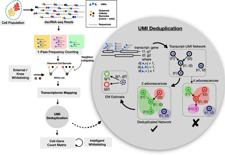
There has been a steady increase in the throughput of single-cell (sc)RNA-seq experiments, facilitating experimental assay of millions of cells. Droplet based scRNA-seq experiments have a large set of gene-ambiguous reads and can commonly account for a quarter of the sequenced data, which stays largely unused by quantification methods. We designed alevin, a fast end-to-end pipeline to process scRNA-seq data, performing cell barcode detection, read mapping, UMI deduplication, gene count estimation, and cell barcode whitelisting. Alevin’s approach to UMI deduplication provides an uncertainty-aware Bayesian model to account for reads that multimap between genes. This addresses the inherent bias in existing tools which discard gene-ambiguous reads and improves the accuracy of gene abundance estimates.

* Mu, Wancen, et al. “Airpart: interpretable statistical models for analyzing allelic imbalance in single-cell datasets.” Bioinformatics 38.10 (2022): 2773-2780.
* Soneson C., Srivastava A., Patro R. & Stadler MB. (2021) “Preprocessing choices affect RNA velocity results for droplet scRNA-seq data.” PLOS Computational Biology. 2021 Jan 11;17(1):e1008585.
* Srivastava A., Malik L., Smith T., Sudbery I. & Patro R. (2019) “Alevin efficiently estimates accurate gene abundances from dscRNA-seq data.” Genome biology. 2019 Dec;20(1):1-6.
* Zhu A., Srivastava A., Ibrahim JG., Patro R. & Love MI. (2019) “Nonparametric expression analysis using inferential replicate counts.” Nucleic Acids Research. 2019 Oct 10;47(18):e105.

3. COMPUTATIONAL METHODS FOR SINGLE-CELL ANALYSES

scRNA-seq data is being generated at an unprecedented pace, and the accurate estimation of gene-level abundances for each cell is a crucial first step in most scRNA-seq analyses. When pre-processing the raw scRNA-seq data to generate a count matrix, care must be taken to account for the potentially large number of multi-mapping locations per read. The sparsity of scRNA-seq data, and the strong 3’ sampling bias, make it even more challenging to disambiguate cases where there is no uniquely mapped read to any of the candidate target genes. We introduced a Bayesian framework for information sharing across cells within a sample or across multiple modalities of data to improve gene quantification estimates for scRNA-seq data.

* Hao, Yuhan, et al. “Dictionary learning for integrative, multimodal and scalable single-cell analysis.” Nature Biotechnology (2023): 1-12.
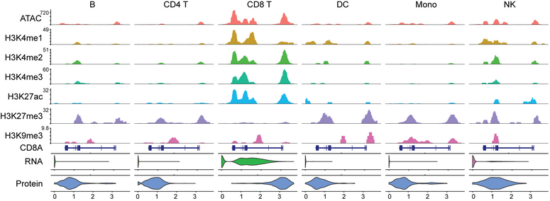
* Zhang, B.*, Srivastava A.*, Mimitou E., Stuart T., Raimondi I., Hao Y., Smibert P. & Satija R. (2021) “Characterizing cellular heterogeneity in chromatin state with scCUT\&Tag-pro.” Nature Biotechnology 40.8 (2022): 1220-1230.
* Stuart T., Srivastava A., Lareau C. & Satija R. (2021) “Single-cell chromatin state analysis with Signac.” Nature Methods (2021): 1-9.
* Srivastava A., Malik L., Sarkar H. & Patro R. (2020) “A Bayesian framework for inter-cellular information sharing improves dscRNA-seq quantification.” Bioinformatics. 2020 Jul 1;36(Supplement\_1):i292-9.

4. INTEGRATED ANALYSES OF THE EPIGENOME TO UNDERSTAND THE MOLECULAR BASIS OF HEMATOPOIETIC MALIGNANCIES

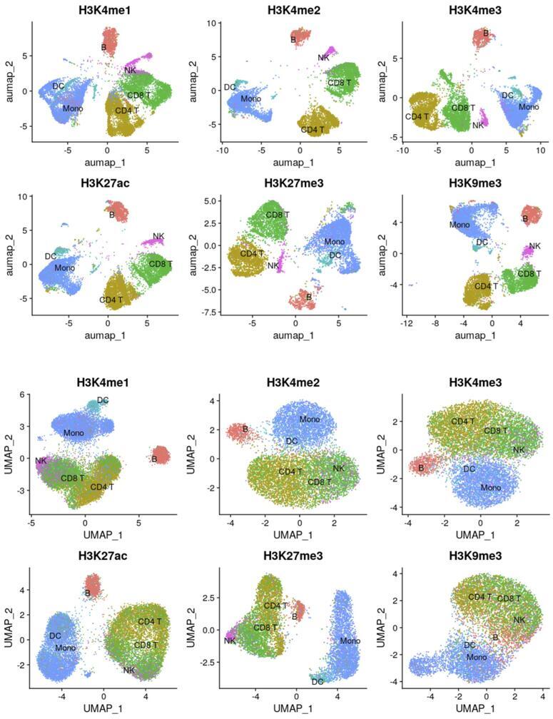
An impaired hematopoietic differentiation process underlies bone marrow malignancies like leukemia, but we still lack the mechanistic understanding of the sequence of regulatory events that misleads the differentiation process. Since epigenomic regulatory patterns are major features of leukemic development, understanding the chromatin dynamics of a failed (malignant) hematopoietic differentiation process can help define the molecular basis of leukemia. A prerequisite to such an understanding is a framework that allows investigation of the progressive changes in the activity of the regulatory elements (RE) during hematopoietic differentiation. Single-cell CUT&Tag (scCUT&Tag) technology is well-suited for such studies as RE activity through histone modification profiles can be investigated in a lineage-specific manner. Using scCUT&Tag we will investigate the RE and progressive changes in their activity during hematopoiesis. First, we will define a multimodal reference mapping framework for mouse hematopoiesis. This framework will allow us to integrate multiple histone modification profiles onto one reference and compare the chromatin states of the RE between a wild type (WT) and mouse model with loss of function in histone methyl transferase (HMT). Second, since HMTs regulate transcription through the interaction network of RE. We will define a chromatin state aware map that dynamically links REs across developmental trajectories. We will use this framework to investigate the changes in the interaction of REs due to HMT loss. Third, since the transcriptional state of a cell emerges from the underlying gene regulatory network (GRN), We will integrate single-cell gene expression data with histone modification profiles and extend it to define a chromatin state aware model of GRN. We will compare the WT and HMT loss experiments and define the differential GRN.

* Srivastava A. (2020) “Integrated analyses of the epigenome to understand the molecular basis of hematopoietic malignancies.”, Project Number: K99CA267677.

Srivastava Lab in the News

Selected Publications

  • Characterizing cellular heterogeneity in chromatin state with scCUT&Tag-pro

    Zhang B, Srivastava A, Mimitou E, Stuart T, Raimondi I, Hao Y, Smibert P, Satija R. Characterizing cellular heterogeneity in chromatin state with scCUT&Tag-pro. Nat Biotechnol. 2022 Aug;40(8):1220-1230. doi: 10.1038/s41587-022-01250-0. Epub 2022 Mar 24. PMID: 35332340; PMCIDPMC9378363.

  • Alevin efficiently estimates accurate gene abundances from dscRNA-seq data

    Srivastava A, Malik L, Smith T, Sudbery I, Patro R. Alevin efficiently estimates accurate gene abundances from dscRNA-seq data. Genome Biol. 2019 Mar 27;20(1):65. doi: 10.1186/s13059-019-1670-y. PMID: 30917859; PMCIDPMC6437997.

  • Alignment and mapping methodology influence transcript abundance estimation

    Srivastava A, Malik L, Sarkar H, Zakeri M, Almodaresi F, Soneson C, Love MI, Kingsford C, Patro R. Alignment and mapping methodology influence transcript abundance estimation. Genome Biol. 2020 Sep 7;21(1):239. doi: 10.1186/s13059-020-02151-8. PMID: 32894187; PMCIDPMC7487471.

  • Dictionary learning for integrative, multimodal and scalable single-cell analysis

    Hao Y, Stuart T, Kowalski MH, Choudhary S, Hoffman P, Hartman A, Srivastava A, Molla G, Madad S, Fernandez-Granda C, Satija R. Dictionary learning for integrative, multimodal and scalable single-cell analysis. Nat Biotechnol. 2023 May 25. doi: 10.1038/s41587-023-01767-y. Epub ahead of print. PMID: 37231261.

  • Alevin-fry unlocks rapid, accurate and memory-frugal quantification of single-cell RNA-seq data

    He D, Zakeri M, Sarkar H, Soneson C, Srivastava A, Patro R. Alevin-fry unlocks rapid, accurate and memory-frugal quantification of single-cell RNA-seq data. Nat Methods. 2022 Mar;19(3):316-322. doi: 10.1038/s41592-022-01408-3. Epub 2022 Mar 11. PMID: 35277707; PMCIDPMC8933848.

View Additional Publications

Continue reading

The Wistar Institute Recruits Dr. Avi Srivastava as Assistant Professor 

PHILADELPHIA—(Sept. 7, 2023)—The Wistar Institute, an international biomedical research leader in cancer, immunology and infectious diseases, is pleased to announce the recruitment of Avi Srivastava, Ph.D., to the Ellen and Ronald Caplan Cancer Center, where he joins Wistar’s Gene Expression and Regulation Program as an Assistant Professor.

A computational biologist, Dr. Srivastava brings expertise in advanced computational methods that can be used to establish powerful predictive research tools in cancer biology. “The opportunity to pursue my research at The Wistar Institute is invaluable,” says Dr. Srivastava. “I’m excited to launch the Srivastava Lab at an institution renowned for its unwavering dedication to cancer research.”

“We welcome Dr. Srivastava with great enthusiasm. His appointment to Wistar demonstrates our continued commitment to expanding our best-in-class research talent,” says Dario Altieri, M.D., Wistar president and CEO, director of the Ellen and Ronald Caplan Cancer Center and the Robert and Penny Fox Distinguished Professor. “Computational biology is set to play a pivotal role in the future of biomedical research, and we are delighted to have Dr. Srivastava contribute his expertise to this critical field here at Wistar.”

###

The Wistar Institute, the first independent, nonprofit biomedical research institute in the United States, marshals the talents of an international team of outstanding scientists through a culture of biomedical collaboration and innovation. Wistar scientists are focused on solving some of the world’s most challenging and important problems in the field of cancer, infectious disease, and immunology. Wistar has been producing groundbreaking advances in world health for more than a century, consistent with its legacy of leadership in biomedical research and a track record of life-saving contributions in immunology and cell biology. wistar.org

Luis J. Montaner, D.V.M., D.Phil

  • Vice President, Scientific Operations

  • Associate Director for Shared Resources, Ellen and Ronald Caplan Cancer Center

  • Herbert Kean, M.D., Family Professor; Director, HIV-1 Immunopathogenesis Laboratory and Leader, HIV Research Program, Vaccine & Immunotherapy Center

  • Immunology, Microenvironment and Metastasis Program, Ellen and Ronald Caplan Cancer Center

  • Scientific Director, Humanized Models of Disease Facility

Montaner studies the mechanisms of disease in HIV-1 infection, cancer, COVID-19, and emerging viral infections (monkeypox, nipah virus), exploring new strategies to boost the natural function of the immune system in order to combat viral-associated disease or cancer progression.

Montaner obtained his D.V.M., Veterinary Medicine from Kansas State University in 1989 and his D.Phil. in Experimental Pathology from University of Oxford, U.K., in 1995. He joined The Wistar Institute in 1995 as an assistant professor and was promoted to professor in 2007. Montaner was named the Herbert Kean, M.D., Family Endowed Chair Professorship in 2015.

View Publications

The Montaner Laboratory

215-898-3934

montanerlab@wistar.org

The Montaner Laboratory

At Wistar, the Montaner laboratory focuses on immune system-based research using laboratory models of virus infection, animal models of infection and/or cancer, and clinical cohort studies to provide a clinic-to-bench research program that informs new strategies to combat HIV and or cancer. The Montaner lab is also a leading center of a Martin Delaney Collaboratory focused on HIV cure-directed research (see beat-hiv.org). Patient- and animal-based collaborative studies extend from Philadelphia across the United States and Puerto Rico, Mexico, Europe, South America, Southern Africa, and Vietnam. Current research focuses on:

  • Identifying new strategies to reverse mechanisms of immunodeficiency caused by viral infection and/or cancer processes via testing new immune-enhancing strategies in patient-based studies (specimens, clinical trials), and animal models (humanized mice models, non-human primates).
  • Exploring new ways to augment HIV-1 control beyond current therapies in order to achieve durable remission and/or permanent control of infection without the need for continued antiretroviral therapy.
  • Understanding the role of targeting myeloid cells in cancer progression.
  • Determining the impact of substance use disorder therapy on immune functionality and HIV reservoir retention in opioid-dependent persons living with HIV.
  • Determining the impact of COVID-19 infection and/or vaccination on immune activation and HIV reservoirs in persons living with HIV.
  • Antiviral discovery strategies based on natural products and small molecule lead optimization.

Watch this video to learn more about HIV cure research.

Staff
  • Research Assistant Professor

    Ian Tietjen, Ph.D.

  • Senior Staff Scientists

    Livio Azzoni, M.D., Ph.D.
    Emmanouil Papasavvas, Ph.D.
    Costin Tomescu, Ph.D.
    Zhe Yuan, Ph.D.

  • Staff Scientist

    Evgenii Tcyganov, Ph.D.

  • Postdoctoral Fellows

    Avishek Bhuniya, Ph.D.
    Khumoekae Richard, Ph.D.

  • Research Assistants

    Yong Cheng
    Matthew Fair
    Colin Hart
    Xiaoshan Jiang
    Brijesh Karanam
    Kai Killebrew
    Adiana Ochoa Ortiz
    Emery Register
    Brian Ross
    Paridhima Sharma
    Guorui Zu

  • Clinical Coordinator

    Ken Lynn, R.N.

  • Clinical Research Assistant

    Giorgio Cocchella

  • BSL-2+ Laboratory Manager

    Jessicamarie Morris

  • Associate Director, HIV Program

    Beth Peterson

  • Administrative Coordinator

    Kathleen Magner

  • Visiting Scientist

    Chantal Emade Nkwele

  • Visiting Scholar

    Fidele Ntie-Kang


Available Positions

Postdoctoral fellow positions are available in the Montaner laboratory, with a focus on cancer and immunotherapy system-based research through a combination of both murine-based models and human translational studies. The preferred candidate is a recent Ph.D. or equivalent with a strong background in immunology, animal models, and molecular/cellular biology. Experience with molecular screening, bioinformatics, microscopy, and flow cytometry is a plus. Individuals eligible for National Research Service Award (NRSA) funding are highly desirable. Motivated candidates are encouraged to submit their CV and references to: montanerlab@wistar.org.

Research

The Montaner lab directs several international teams and advances basic translational research focused on immunology, infectious disease and cancer. The lab has expertise on human immunology (innate response), HIV cure-directed efforts, cancer immunotherapy, and clinical trials.

  • BEAT-HIV Delaney Collaboratory to Cure HIV-1 Infection by Combination Immunotherapy

    In 2021, based on the research progress made, the BEAT-HIV Collaboratory received a second, five-year, $29.15 million award and was joined by a third principal investigator Robert Siliciano, M.D., Ph.D., from Johns Hopkins University in addition to co-principal investigators Luis J. Montaner, D.V.M., D.Phil., from Wistar, and James L. Riley, Ph.D., from the University of Pennsylvania. The new award is one of 10 grants funded in 2021 by the National Institutes of Health’s (NIH) “Martin Delaney Collaboratories to Cure HIV” initiative to a highly-select group of U.S.-led teams charged with advancing global efforts to develop a cure for HIV. Building on the success of the initial award made in 2016, when the NIH awarded nearly $23 million to the BEAT-HIV Delaney Collaboratory to Cure HIV-1 Infection by Combination Immunotherapy. The most recent cycle of funding does not include a clinical trial.

    The BEAT-HIV Delaney Collaboratory is a partnership of more than100 leading HIV investigators, from academia to industry partners working with community-based organization Philadelphia FIGHT, to test combinations of several novel immunotherapies under new preclinical research. The new cycle of BEAT-HIV funding established three research goals, based on goals outlined in the NIH program announcement:

    • Understand the basic mechanisms underlying persistence of the viral reservoir during ART, what cell populations contribute to rebound after treatment interruption, and the role played by host-related factors.
    • Develop strategies to achieve durable suppression of HIV replication in the absence of ART. Capitalizing on advances in clinical research on bNAbs, BEAT-HIV researchers will test synthetic DNA technology to better deliver the genetic blueprint for the body to make different specific bNAbs simultaneously. An additional approach will be to boost natural killer and T cell responses to achieve long-term viral suppression. Eventually, the two strategies will be combined to maximize long-term control potential.
    • Develop new approaches to eradicate the HIV reservoir. Strategies to be tested include novel drugs able to reactivate latent HIV hiding in the immune cells, combined with CAR-T cell approaches designed to change a person’s killer T cells to make them able to find infected cells more efficiently. In addition, researchers will apply a technology called mRNA-LNP to make cells resistant to HIV. The ultimate goal is to identify which approaches have the best potential and test them in combination to achieve complete HIV eradication.
    BEAT-HIV2 CLINICAL STUDIES – COMPLETE

    We completed enrollment in each HIV cure-directed clinical trial funded under the BEAT-HIV umbrella from 2016-2021.

    The first study was to determine if treatment with pegylated interferon alpha 2b (peg-IFN-α2b) together with neutralizing antibodies 3BNC117+10-1074 will result in a reduction of viral rebound and reduction in the amount of latent HIV DNA in peripheral blood cells and tissues of individuals with chronic HIV infection upon an antiretroviral treatment (ART) interruption. By measuring the changes in viral rebound after ART interruption as a surrogate measure of the latent reservoir and immune control, the study will establish if this combined immunotherapy strategy should be considered as a component of future viral eradication strategies. Visit ClinicalTrials.gov under NCT03588715 for more information. Visit ClinicalTrials.gov under NCT03588715 for more information. Data analysis is underway.

    The second study tested a clinical strategy that combined two gene therapy vectors to genetically modify T cells purified from study participants using the chimeric antigen receptor (CAR) technology to make these cells highly specific in recognizing HIV-infected cells. In addition, these T cells will be made HIV-resistant by using Zinc-finger Nucleases (ZFNs) that target CCR5, an HIV entry molecule. As a result of these genetic modifications, immune cells will be rendered specific in their killing capacity while also resistant against HIV infection, which is expected to enhance their intrinsic ability to clear HIV-infected cells and result in durable viral suppression after suspension of the antiretroviral therapy. To learn more about this clinical trial, visit ClinicalTrials.gov (NCT03617198). Data analysis is underway.

    HOME-BASED VIRAL LOAD TESTING DEVICE – ENROLLMENT BY INVITATION

    BEAT-HIV investigators are partnering with Merck, Inc. and Tasso, Inc. to assess the reliability and acceptability of a home-based viral load testing device. The micro-blood collection device was previously tested with participants enrolled in each of the two BEAT-HIV clinical trials described above.

    HIV cure-directed clinical trials often include an analytic treatment interruption (ATI, learn more about that here). ATIs require frequent clinic visits to monitor participants’ viral load to keep it within study safety guidelines. The COVID epidemic changed the research landscape, including a stated desire among study participants and community advocates to reduce the number of study visits (and potential exposure to COVID-19 during transit and at the clinic).

    The device being tested could potentially reduce the number of required clinic visits for blood collection, but only if the home-based viral load device test works as well as standard lab-based viral load testing that can now only be done at the clinic. Just as important is to determine how people living with HIV feel about the device and the process for returning it to the lab by mail or courier, how comfortable they are with using the home-based device, and if they have any other concerns.

    The home-based viral load testing device is currently being offered by invitation to participants resuming antiretroviral therapy. At this time, the home-based viral load testing device is available within an experimental framework to determine if: 1) the device is acceptable to participants and 2) if the device provides results that are comparable to those available with traditional clinic- or lab-based viral load testing.

  • AMOHI Consortia

    OPIOIDS AND HIV: PHILADELPHIA-BASED STUDY TO DEFINE HOST FACTORS DRIVING HIV INFECTION AND IMPACTING OPTIMAL MANAGEMENT OF OPIOID USE DISORDER

    Pennsylvania is at the center of the opioid and HIV epidemic in the U.S., leading in both new HIV infections associated with persons who inject drugs (PWIDs) and in overdose deaths due to lack of optimal management of opioid drug addiction. In 2019, the federal government identified Philadelphia County as one of 48 counties responsible for >50% of new HIV diagnoses in the U.S. New infections in Philadelphia have been linked to an increase in HIV transmission from PWIDs.

    Pennsylvania also ranks third nationwide for deaths due to injection-drug overdose. Management of opioid addiction in persons living with HIV who are already in care for their HIV disease can be achieved by use of medications for opioid use disorder (MOUDs), including methadone, buprenorphine, or naltrexone. Even though these medications can impact immune activation on their own, what remains unknown is how the choice of methadone, buprenorphine, or naltrexone could affect the immune recovery and therefore, the future health of persons taking medications for their HIV disease and MOUD.

    The Montaner lab and collaborators in Prevention Point Philadelphia, University of Pennsylvania, and Philadelphia FIGHT are investigating the host factors driving new HIV infections in persons who inject drugs and the impact of MOUDs on immune recovery after HIV suppression by antiretroviral therapy (ART).

    PHILADELPHIA MECHANISTIC STUDY – CURRENTLY ENROLLING

    We have established clinical access to target populations in Philadelphia. We will use this clinical infrastructure (mobile units and multi-city clinical sites) to collect substance use/behavior (questionnaires) and biological data (blood samples) from target PWIDs who are at high risk of HIV infection, as well as HIV-infected PWIDs who are currently under medications for opioid use disorder. In collaboration with University of Pennsylvania, Jonathan Lax Treatment Center, the Icahn School of Medicine at Mount Sinai, and Prevention Point Philadelphia among others, we will conduct a mechanistic study to determine the levels of inflammation and innate immune activation in depressed and non-depressed PWIDs, and if levels of immune activation can impact HIV susceptibility ex vivo. We will also define levels of immune activation associated with comorbidities (if elevated) in HIV-infected PWIDs who are stably suppressed under ART and taking either methadone, buprenorphine, or naltrexone to evaluate the impact of long-term opioid receptor stimulation or blockage with MOUDs on immune reconstitution in persons living with HIV. The long-term impact of this study is to investigate novel factors that can be targeted for HIV prevention in PWIDs and/or how best to manage opioid drug addiction in HIV-infected persons to ensure the best immune recovery after HIV therapy able to reduce added comorbidities in the future while increasing overall survival.

    AMOHI CLINICAL TRIAL (VIETNAM)

    NIH-funded projects trial will provide clinical evidence to investigate the link between retention of chronic immune activation in HIV-1-infected opioid users receiving medication for opioid use disorder (MOUD) combined with antiretroviral therapy (ART) and starting on methadone maintenance, when compared to naltrexone or buprenorphine.

    The Montaner lab leads an international team composed of investigators from the U.S., Vietnam, and France, in collaboration with the Vietnam Ministry of Health, University of Pennsylvania, IMEA (a French-led initiative to expand access to HIV/hepatitis prevention and treatment services), the Pasteur Institute, and industry partners Alkermes, plc and Rusan. The goal of this three-arm randomized trial is to evaluate the impact of long-term opioid receptor stimulation or blockage with MOUDs on immune reconstitution in HIV-infected people who inject drugs and are initiating ART. Early preliminary data suggest that chronic opioid receptor engagement by an opioid receptor agonist while on ART may result in increased immune activation and inflammation associated with increased levels of persistent HIV, when compared to a full opioid receptor antagonist. To verify this hypothesis, the study will assess recovery outcomes and adherence to therapy 48 weeks after initiation of ART in 225 participants with OUD who receive either methadone (opioid receptor agonist), extended-release naltrexone (antagonist) or buprenorphine (partial agonist).

  • Coronavirus Small Molecule Drug Discovery Program

    Working with medicinal chemists and SARS-CoV-2 wildtype and variants in Wistar BSL-3 facilities, we aim to develop novel combination therapies against COVID-19 based on small molecules:

    • Small molecules that amplify the natural interferon-based host resistance already shown in early therapy trials to reverse detrimental COVID-19 disease progression. We have found small molecules that enhance the natural antiviral responses mediated by existing type I interferons without inducing further inflammation damage on their own. Once developed, this novel therapy is also expected to be applicable for use with antivirals in future viral outbreaks.
    • Small molecules that inhibit viral spread by directly blocking SARS-CoV-2 Spike protein (S) interactions with the ACE2 receptor and/or inhibiting the activity of the viral MProtease enzyme (Mpro or 3CLpro) in infected cells. We have identified small molecules that disrupt the interaction of the S protein with its human ACE2 receptor, thereby inhibiting viral entry; and small molecules that disrupt the MPro enzyme. MPro is a papain-like cysteine protease essential for processing the polyproteins that are translated from the viral RNA. Mpro can process at least 11 cleavage sites on the large polyprotein 1ab, the multifunctional protein involved in the transcription and replication of the viral RNAs. Inhibiting the activity of this enzyme would block viral replication. Because no human proteases with a similar cleavage specificity are known, such inhibitors are less likely to be toxic. We will also take advantage of the large and unique natural product and synthetic libraries available at Wistar to identify added lead molecules. We will validate identified hits using established SARS-CoV-2 Spike pseudo-virus systems as well as a live viral challenge model.

    Development of these therapeutics, to be used in combination at onset of symptoms or in those at high risk of developing symptoms, is expected to limit viral infection, preserve lung tissue integrity, and prevent progression towards a cytokine release (“storm”) syndrome associated with mortality.

  • Myeloid Cells and Cancer Progression

    Myeloid cells are critical components of the tumor microenvironment. Under physiological conditions these cells are comprised of mature terminally differentiated cells: polymorphonuclear neutrophils (PMN) and other granulocytes; macrophages (MΦ); and dendritic cells (DCs). In cancer, the myeloid compartment is dramatically affected, which is now considered one of the major immunological hallmarks of cancer. Tumor-bearing (TB) hosts accumulate immunosuppressive MΦ, DCs that are ineffective in inducing potent immune responses. The prominent change in the myeloid compartment in cancer is the expansion of pathologically activated immature myeloid cells with a potent ability to suppress immune responses — myeloid-derived suppressor cells (MDSC). In TB mice, the total population of MDSC consists of three groups of cells: pathologically activated neutrophils (PMN-MDSC) are the most abundant (>75%); pathologically activated monocytes (M-MDSC) are less abundant (<20%); and early myeloid precursors represent a small (<5%) population. The current view considers changes in myeloid cells separately, with different mechanisms applied to the different cell types. The gap in our knowledge is how these different myeloid cells can interact with each other in TB hosts. We are investigating the bridge between different populations of myeloid cells in cancer and how they orchestrate their abnormal function. The ultimate goal of this project is not only to better understand the mechanism regulating myeloid cell function in cancer, but to develop novel approaches for regulation of immune responses in cancer.

  • Development of Novel Small-molecule Rb Protein Modulator as Cancer Immunotherapy

    According to National Cancer Institute (NCI) statistics, ovarian cancer represents 1.3% of all cancers, and more than 21,000 women are diagnosed every year in the U.S. An estimated one woman in 75 will develop ovarian cancer during her lifetime. Although many therapeutic approaches have been tested, including surgery, radiation, chemotherapy, and immunotherapy, ovarian cancer remains extremely difficult to treat, and novel therapeutic approaches are needed. This project, funded in part by the Department of Defense, is based on therapeutic strategies that can modulate myeloid cell apoptosis resulting in an increase of anti-tumor immune responses.

  • Humanized Mouse Program: Cancer and Infectious Disease

    Absent direct clinical trials in humans, animal models of HIV infection are the best platform to explore novel pre-clinical anti-HIV strategies. Animal models for HIV infection include nonhuman primates and humanized mice. Humanized mice have emerged as a model able to be used for high-volume screening, yet the suboptimal immune differentiation that occurs has raised concern on the ability of this model to fully reflect all aspects of an immune response otherwise present in humans. The humanized mouse system has been developed to model HIV infection in humans, response to antiretroviral therapy (ART) and novel cure interventions, as well as study cancer immunotherapy in patient-derived xenograft models. A new WistarHu mice platform has been developed to support cancer immunotherapy and Wistar-based discovery of strategies against HIV based on assessing changes in viral measures on ART or effects on viral load rebound after ART interruption (Analytical Therapy Interruption, ATI). In support of this new platform, we have established ART formulations, HIV infection, HIV suppression, and characterized changes on immune reconstitution, persistent HIV measures, microbial translocation after ART and during an ATI. This platform is currently applied toward discovery and collaborative work.

  • HIV-1 Patient Partnership Program: Basic Research and HIV Social Science

    With long-standing commitment from Philadelphia FIGHT and the University of Pennsylvania along with the Robert I. Jacobs Fund of The Philadelphia Foundation, the HIV-1 Patient Partnership Program was established to provide clinical material for basic research and to sponsor the Jonathan Lax Memorial Lecture (also supported by Henry S. Miller, Jr. and Ken Nimblett). Research with clinical material obtained from this program is focused on mechanisms of AIDS immunopathology. This collaborative link between our research team and over 6000 HIV-1 patients in the Philadelphia region has led to the largest HIV Cure clinical trial to date — the BEAT-HIV Study.

    The HIV-1 patient-partnership program involving participants in research is based at Philadelphia FIGHT (a community-based HIV-1 primary care provider) and Prevention Point Philadelphia. Our partnership with each community-based organization strives to develop trusted relationships and maintain meaningful, bi-directional lines of communication between investigators and communities most affected by HIV and substance use disorder. The primary objective of our community engagement strategy is to ensure communities have a clear understanding of a) the research being implemented, whether HIV-cure directed or other HIV, COVID, or monkeypox (MPOX) research, b) the stage of the research (including realistic expectations around HIV cure science), and c) how interested individuals can participate in and support our research agenda.

    Social Science was added to our HIV cure research agenda to enhance both our preclinical and community engagement efforts. The Social Sciences Initiative assesses the acceptability of HIV cure interventions under development and conducts empirical ethics research related to HIV cure. Working in close collaboration, BEAT-HIV investigators and community stakeholders have developed a robust agenda of educational activities, community-based projects, and basic/clinical research designed to ensure comprehensive understanding and to provide guidance on the ethical conduct of HIV cure-directed research.

    Above all, the Montaner laboratory makes its research accountable to our study participants and other stakeholders through community advisory board (CAB) review and community representation on Data Safety Monitoring Boards for clinical trials when active. In addition, we provide community-focused research presentations at Philadelphia’s AIDS Education Month, the annual Lax Lecture, and other community events, so that community members and other interested individuals are informed about the outcomes of patient-supported research.

    The Jonathan Lax Lecture honors the memory of Jonathan Lax, a businessman, inventor, teacher, and one of the best-known AIDS activists in Philadelphia’s community-based clinical research network, where he volunteered with many groups to try and speed the drug approval process. He left funds to start a clinic — today called the Jonathan Lax Center — that is now the largest provider of AIDS care in Philadelphia, independent of a patient’s ability to pay. The Lax Lecture is a public lecture held in June of each year at The Wistar Institute, where leading international HIV scientists interact with local researchers, clinicians, and patient advocates. Previous speakers include Gates Foundation HIV Frontiers and Biotechnology Accelerator head Mike McCune, Partners in Health founder and Harvard professor Paul Farmer, Project Inform founder Martin Delaney, and 2008 Nobel Laureate Françoise Barré-Sinoussi. In 2021, the Lax Lecture celebrated its 25th year by honoring Anthony Fauci for his dedication to serving people living with HIV – the first person to receive this honor twice.

View a list of previous projects pursued by the Montaner lab.

Global Health & Partnerships

The Wistar Institute fosters a local and global community that is unified by bold scientific thinking, leadership, and a collaborative spirit. Thought leaders from nonprofits, healthcare, pharmaceutical and biotechnology companies, governments and other agencies of influence choose to work collaboratively with Wistar scientists to accelerate the creation of new therapies for patients worldwide. We have connections in locations here.

Staff
  • Research Assistant Professor

    Ian Tietjen, Ph.D.

  • Senior Staff Scientists

    Livio Azzoni, M.D., Ph.D.
    Emmanouil Papasavvas, Ph.D.
    Costin Tomescu, Ph.D.
    Zhe Yuan, Ph.D.

  • Staff Scientist

    Evgenii Tcyganov, Ph.D.

  • Postdoctoral Fellows

    Avishek Bhuniya, Ph.D.
    Khumoekae Richard, Ph.D.

  • Research Assistants

    Yong Cheng
    Matthew Fair
    Colin Hart
    Xiaoshan Jiang
    Brijesh Karanam
    Kai Killebrew
    Adiana Ochoa Ortiz
    Emery Register
    Brian Ross
    Paridhima Sharma
    Guorui Zu

  • Clinical Coordinator

    Ken Lynn, R.N.

  • Clinical Research Assistant

    Giorgio Cocchella

  • BSL-2+ Laboratory Manager

    Jessicamarie Morris

  • Associate Director, HIV Program

    Beth Peterson

  • Administrative Coordinator

    Kathleen Magner

  • Visiting Scientist

    Chantal Emade Nkwele

  • Visiting Scholar

    Fidele Ntie-Kang


Available Positions

Postdoctoral fellow positions are available in the Montaner laboratory, with a focus on cancer and immunotherapy system-based research through a combination of both murine-based models and human translational studies. The preferred candidate is a recent Ph.D. or equivalent with a strong background in immunology, animal models, and molecular/cellular biology. Experience with molecular screening, bioinformatics, microscopy, and flow cytometry is a plus. Individuals eligible for National Research Service Award (NRSA) funding are highly desirable. Motivated candidates are encouraged to submit their CV and references to: montanerlab@wistar.org.

Staff Highlight: Ian Tietjen, Ph.D., Focuses on Bringing Traditional Medicine into Modern Research

Ian Tietjen, Ph.D., focuses on mechanisms of viral pathogenesis, and on drug discovery and development. He uses cell biology, genetics, and high-throughput chemical screening techniques to investigate the molecular properties of HIV reservoirs in addition to influenza, coronavirus, and nipah virus.

Tietjen collaborates with local communities, medicinal plant healers, and other knowledge keepers to sustainably and ethically document and determine the bioactivities of traditional medicines used in Southern Africa, Canada, and elsewhere.

Tietjen joined Wistar as a research assistant professor in the HIV Research Program in January 2020 and he is the head of the Small Molecule Discovery and Pharmacognosy Group. He was previously an assistant professor in the Faculty of Health Sciences in Vancouver, Canada, and has worked as a group leader in Molecular and Cellular Biology at Cardiome Pharma Corp. and a senior scientist at Xenon Pharmaceuticals.

Click here to see Tietjen’s select publications.

SMALL MOLECULE DISCOVERY AND PHARMACOGNOSY GROUP

The Small Molecule Discovery and Pharmacognosy Group works with researchers, traditional healers, and other knowledge keepers who are interested in identifying and elucidating the molecular and biomedical properties of naturally produced chemical compounds and medicinal plants. The group primarily focuses on potential therapies for HIV, coronaviruses, influenza, and other infectious pathogens but also supports studies for cancer, metabolic diseases, and other illnesses. We provide assay development, laboratory training and instruction, and community engagement expertise to meaningfully work with local and Indigenous communities with traditional medicinal knowledge.

Individuals interested in working with the Small Molecule Discovery and Pharmacognosy Group can contact Ian Tietjen for more information at itietjen@wistar.org.

The mission of the BEAT-HIV Collaboratory is the define the most effective way to combine immunotherapy regimens to cure HIV.

Learn more about BEAT-HIV

Lab Events and Photos

2016 Staff Lunch

Montaner Lab in the News

Selected Publications

  • Gene Expression Profiling Informs HPV Cervical Histopathology but Not Recurrence/Relapse After LEEP in ART-Suppressed HIV+HPV+ Women.

    Papasavvas, E., Kossenkov, A.V., Azzoni, L., Zetola, N.M., Mackiewicz, A., Ross, B.N., Fair, M., Vadrevu, S., Ramogola-Masire, D., Sanne, I., Firnhaber, C., Montaner, L.J. “Gene Expression Profiling Informs HPV Cervical Histopathology but Not Recurrence/Relapse After LEEP in ART-Suppressed HIV+HPV+ Women.” Carcinogenesis. 2019 Apr 29;40(2):225-233. doi: 10.1093/carcin/bgy149.

  • Intact HIV reservoir estimated by the intact proviral DNA assay correlates with levels of total and integrated DNA in the blood during suppressive antiretroviral therapy.

    Papasavvas, E., Azzoni, L., Ross, B.N., Fair, M., Yuan, Z., Gyampoh, K., Mackiewicz, A., Sciorillo, A.C., Paggliuzza, A., Lada, S.M., Guoxin, W., Goh, S.L., Bahnck-Teets, C., Holder, D.J., Zuck, P.D., Damra, M., Lynn, K.M., Tebas, P., Mounzer, K., Kostman, J.R., Abdel-Mohsen, M., Richman, D., Chomont, N., Howell, B.J., Montaner, L.J. “Intact HIV reservoir estimated by the intact proviral DNA assay correlates with levels of total and integrated DNA in the blood during suppressive antiretroviral therapy.” Clin Infect Dis. 2020 Jun 18;ciaa809. doi: 10.1093/cid/ciaa809. Online ahead of print.

  • Cytokine storm and leukocyte changes in mild versus severe SARS-CoV-2 infection: Review of 3939 COVID-19 patients in China and emerging pathogenesis and therapy concept.

    Wang, J., Jiang, M., Chen, X., Montaner, L.J. “Cytokine storm and leukocyte changes in mild versus severe SARS-CoV-2 infection: Review of 3939 COVID-19 patients in China and emerging pathogenesis and therapy concept.” J Leukoc Biol. 2020 Jul;108(1):17-41. doi: 10.1002/JLB.3COVR0520-272R.

  • Recommendations for measuring HIV reservoir size in cure-directed clinical trials.

    Abdel-Mohsen, M., Richman, D., Siliciano, R.F., Nussenzweig, M.C., Howell, B., Martinez-Picado, J., Chomont, N., Bar, K.J., Yu, X.G., Lichterfeld, M., Alcami, J., Hazuda, D., Bushman, F., Siliciano, J., Betts, M.R., Spivak, A.M., Planelles, V., Hahn, B.H., Smith, D.M., Ho, Y., Buzon, M.J., Gaebler, C., Paiardini, M., Li, Q., Estes, J.D., Hope, T.J., Kostman, J., Mounzer, K., Caskey, M., Fox, L., Frank, L., Riley, J.L., Tebas, P., Montaner, L.J., BEATHIV Delaney Collaboratory to Cure HIV-1 infection “Recommendations for measuring HIV reservoir size in cure-directed clinical trials.” Nat Med. 2020 Sep;26(9):1339-1350. doi: 10.1038/s41591-020-1022-1.

  • Effect of Opioid Use on Immune Activation and HIV Persistence on ART.

    Azzoni, L., Metzger, D., Montaner, L.J. “Effect of Opioid Use on Immune Activation and HIV Persistence on ART.” J Neuroimmune Pharmacol. 2020 Dec;15(4):643-657. doi: 10.1007/s11481-020-09959-y.

View Additional Publications

Continue reading

Bin Tian, Ph.D.

  • Professor and Program Co-Leader, Gene Expression and Regulation Program, Ellen and Ronald Caplan Cancer Center

  • Co-director, Center for Systems & Computational Biology

Tian is a molecular systems biologist whose research is focused on understanding how gene expression is regulated at the RNA level. His lab was among the first to discover the widespread nature of alternative polyadenylation (APA) using bioinformatic and genomic approaches. They have also revealed multiple molecular mechanisms that regulate APA and cellular functions of APA isoforms in a number of biological systems.

Tian received his B.S. degree in biochemistry from East China University of Science and Technology and his Ph.D. degree in molecular biology from Rutgers Biomedical and Health Sciences (formerly UMDNJ). He was a postdoctoral fellow in bioinformatics and genomics at Johnson & Johnson Pharmaceutical Research & Development in La Jolla, California. In 2003, he established his research group at Rutgers New Jersey Medical School, where he rose through the ranks and became a tenured professor in 2014. Tian joined The Wistar Institute in 2020.

View Publications

The Tian Laboratory

215-898-3922

btian@wistar.org

The Tian Laboratory

Expression of the genetic code, from DNA to protein, can be regulated at different stages, much of which takes place after RNA is made. The Tian lab studies RNA biology using a variety of approaches including functional genomics, computational biology, and molecular and cellular biology. They have contributed important knowledge on the mechanisms and consequences of alternative polyadenylation (APA) in development and disease.

Staff
  • Visiting Scientist

    Wei Chun Chen, Ph.D.

  • Postdoctoral Fellows

    Yange Cui, Ph.D.
    Luyang Wang, Ph.D.
    Qiang Zhang, Ph.D.

  • Graduate Student

    Yuxi Ai (UPenn, Biochemistry and Molecular Biophysics)

  • Research Assistant

    Qingbao Ding


Available Positions

Multiple graduate student and postdoctoral positions are available in the Tian lab. Motivated candidates interested in experimental studies, or computational research or both are encouraged to inquire about the positions by contacting Dr. Bin Tian, btian@wistar.org.

Research

Functional Genomics of Cleavage and Polyadenylation

In eukaryotes, almost all protein-coding mRNAs and long non-coding RNAs (lncRNAs) transcribed by RNA polymerase II employ cleavage and polyadenylation (CPA) for 3’ end maturation. CPA is also coupled with termination of transcription. A gene can have multiple cleavage and polyadenylation sites (PASs), resulting in mRNA isoforms with different coding sequences and/or 3’ untranslated regions (3’UTRs), a phenomenon known as alternative cleavage and polyadenylation (APA). The Tian lab is using novel sequencing methods to identify PASs in major model species to understand the evolution of APA. They are also examining APA dynamics in different cells under various pathological and physiological conditions using single cell-based transcriptome data. The long-term goal is to develop an APA code for APA regulation.

3’UTR-mediated Spatial and Temporal Control of mRNA Metabolism

The 3’UTR plays regulatory roles in mRNA metabolism, including mRNA decay, translation, and localization. Sequence and structural motifs embedded in 3’UTRs contribute to 3’UTR functions through interactions with their cognate RNA binding proteins (RBPs), microRNAs (miRNAs), or lncRNAs. The Tian lab recently reported widespread translation-independent endoplasmic reticulum association (TiERA) of mRNAs, in which 3’UTRs play an important role. They are now using cell biology and genomic techniques to examine the underlying mechanism of TiERA. In addition, they are analyzing how ER stress regulates 3’UTR-mediated post-transcriptional control.

mRNA Isoform Regulation in Immunity

The Tian lab recently reported widespread transcript shortening in secretory cell differentiation. The phenomenon, named secretion-coupled APA (SCAP), was observed in multiple professional secretory cells. They are now studying SCAP in B cell differentiation to plasma cells, which is critical for humoral immunity. In addition, activation of other immune cells, such as T cells, monocytes, and macrophages, also involves mRNA isoform changes, although the underlying mechanisms are not the same. The Tian lab is examing regulators involved in mRNA isoform changes in different types of immune cells and key effectors involved in immune reponses. They are also pursuing novel therapeutics to modulate immunity via perturbation of mRNA isoform biogenesis and metabolism.

Transcriptional Termination in Cancer Cells

Dysregulation of 3’ end processing has been shown in multiple cancers. The Tian lab is studying certain cancer cells displaying unusal APA isoform profiles that indicate reliance on 3’ end processing activities for cell survival. In addition, they are studying how transcriptional termination is connected with genome intergrity and APA-associated neoantigens in cancer cells. These studies can lead to novel therapeutic modalities for cancer.


Databases and Software

PolyA_DB is a web-based database created by the Tian lab for comprehensive cataloging of pre-mRNA cleavage and polyadenylation (polyA) sites in multiple species. Learn More.

APAlyzer is a bioinformatics program developed by the Tian lab for the analysis of APA isoform expression changes by using RNA-seq data. Learn More.

MAAPER is a bioinformatics program co-developed by the Tian lab and the Li lab at Rutgers University for APA isoform expression analysis by using 3’ end-biased RNA-seq data from bulk samples or single cells.

Tian Lab in the News

Selected Publications

  • Alternative 3′ UTRs Play A Widespread Role In Translation-independent mRNA Association With The Endoplasmic Reticulum.

    Cheng, L.C., Zheng, D., Zhang, Q., Guvenek, A., Cheng, H., Tian, B. “Alternative 3’ UTRs Play A Widespread Role In Translation-independent mRNA Association With The Endoplasmic Reticulum.” Cell Rep. 2021 Jul 20;36(3):109407. doi: 10.1016/j.celrep.2021.109407.

  • Widespread Transcript Shortening Through Alternative Polyadenylation in Secretory Cell Differentiation.

    Cheng, L.C., Zheng, D., Baljinnyam, E., Sun, F., Ogami, K., Yeung, P.L., Hoque, M., Lu, C., Manley, J.L., Tian, B. “Widespread Transcript Shortening Through Alternative Polyadenylation in Secretory Cell Differentiation.” Nat Commun. 2020 Jun 23;11(1):3182. doi: 10.1038/s41467-020-16959-2.

  • Regulation Of Intronic Polyadenylation By PCF11 Impacts mRNA Expression Of Long Genes.

    Wang, R., Zheng, D., Wei, L., Ding, Q., Tian, B. “Regulation Of Intronic Polyadenylation By PCF11 Impacts mRNA Expression Of Long Genes.” Cell Rep. 2019, Mar 5;26(10):2766-2778. doi: 10.1016/j.celrep.2019.02.049.

  • A Compendium Of Conserved Cleavage And Polyadenylation Events In Mammalian Genes.

    Wang, R., Zheng, D., Yehia, G., Tian, B. “A Compendium Of Conserved Cleavage And Polyadenylation Events In Mammalian Genes.” Genome Res. 2018 Oct;28(10):1427-1441. doi: 10.1101/gr.237826.118. Epub 2018 Aug 24.

  • Cellular Stress Alters 3’UTR Landscape Through Alternative Polyadenylation And Isoform-specific Degradation.

    Zheng D., Wang, R., Ding, Q., Wang, T., Xie, B., Wei, L., Zhong, Z., Tian, B. “Cellular Stress Alters 3’UTR Landscape Through Alternative Polyadenylation And Isoform-specific Degradation.” Nat Commun. 2018 Jun 11;9(1):2268. doi: 10.1038/s41467-018-04730-7.

View Additional Publications

Continue reading

Meenhard Herlyn, D.V.M., D.Sc.

  • Professor, Molecular and Cellular Oncogenesis Program, Ellen and Ronald Caplan Cancer Center

  • Director, The Wistar Institute Melanoma Research Center

Herlyn studies the normal and malignant tissue environment to develop rational approaches to cancer therapy, with a focus on melanoma, the most aggressive form of skin cancer.

Born and educated in Germany, Herlyn received his D.V.M. at the University of Veterinary Medicine, Hanover in 1970 and went on to receive a D.Sc. in medical microbiology at the University of Munich in 1976. He came to The Wistar Institute as an associate scientist in 1976, where he worked in the emerging field of monoclonal antibodies, a technology that formed the basis of a portion of today’s new targeted therapeutics. In 1981, Herlyn became an assistant professor and established a laboratory that is, today, one of the best-known research groups on the study of melanoma biology.

View Publications

The Herlyn Laboratory

215-495-6883

jkohn@wistar.org

The Herlyn Laboratory

The Herlyn laboratory at The Wistar Institute focuses on the biology that underlies melanoma. His efforts have pioneered the use of the three-dimensional “artificial skin” cultures to study the behavior of both tumor and normal cells that sustain tumor growth, a system known as the tumor microenvironment. The Herlyn Laboratory has transformed the scientific understanding of stem cells as they relate to cancer, and their work on the networks of signaling pathways in melanoma has formed the basis of numerous therapies now in clinical trials or recently approved.

Staff
  • Staff Scientists

    Haiyin Li, Ph.D.
    Haiwei Mou, Ph.D.
    Eric Ramirez-Salazar, D.Sc.

  • Postdoctoral Fellows

    Gatha Thacker, Ph.D.
    Qiuxian Zheng, Ph.D.

  • Predoctoral Trainee

    Jessica Kaster

  • Bioinformatics Technician

    Yeqing Chen, M.S.

  • PDX Core Manager

    Monzy Thomas, Ph.D.

  • Wistar Research Assistants

    Ling Li, M.D.
    Min Xiao, M.S.

  • Research Assistants

    Finley Medina
    Veronika Yakovishina
    Maggie Dunne

  • Undergraduate Students (UPenn)

    Hannah Hamdani
    Vincent Ni
    Elaine Tanel

  • Technician

    Abdiel Mandella Reynolds

  • Lab Coordinator

    Jessica Kohn

Resources

CELL LINES

Due to the high demand for our cell lines, the Herlyn lab is pleased to collaborate with Rockland Immunochemicals Inc. for distribution.

Please contact Rockland customer service directly about acquiring our melanoma cell lines.

For help selecting lines, see the mutational chart below or search using Rockland’s website. For any questions, concerns, help, or other issues, please email Min Xiao at Wistar or contact customer service at Rockland.

View Cell Lines

STR PROFILES

Cell authentication has received considerable attention recently as more and more reports of cell line cross contaminations and misidentifications have come to light. As such, in 2008, we implemented and routinely perform Short tandem repeat (STR) profiling using AmpFlSTR® Identifiler® PCR Amplification Kit (Catalog Number 4322288) by Life Technologies which uses loci consistent with all major worldwide STR standards. PCR amplification and STR allele separation and sizing is performed by the Wistar Genomics Facility. Profile interpretation is performed in the Herlyn lab by interrogating the resulting DNA fingerprint to our internal database which includes over 1000 fingerprints, primarily Wistar Melanoma but also commonly used cell lines such as HeLa and 293T cells. The STR profile is provided here as a reference comparison to your results.

For additional inquiries, please email Min Xiao.

Download STR Profiles

Staff
  • Staff Scientists

    Haiyin Li, Ph.D.
    Haiwei Mou, Ph.D.
    Eric Ramirez-Salazar, D.Sc.

  • Postdoctoral Fellows

    Gatha Thacker, Ph.D.
    Qiuxian Zheng, Ph.D.

  • Predoctoral Trainee

    Jessica Kaster

  • Bioinformatics Technician

    Yeqing Chen, M.S.

  • PDX Core Manager

    Monzy Thomas, Ph.D.

  • Wistar Research Assistants

    Ling Li, M.D.
    Min Xiao, M.S.

  • Research Assistants

    Finley Medina
    Veronika Yakovishina
    Maggie Dunne

  • Undergraduate Students (UPenn)

    Hannah Hamdani
    Vincent Ni
    Elaine Tanel

  • Technician

    Abdiel Mandella Reynolds

  • Lab Coordinator

    Jessica Kohn

PDX

The laboratory has generated more than 500 patient-derived xenografts (PDX), which have been extensively characterized. A selection of the PDX is available through Envigo.

ADDITIONAL RESOURCES

For inquiries regarding any of the following techniques or resources, please email Jessica Kohn to be put in touch with the appropriate lab representative.

Cell Culture Techniques

Stem Cells


Research

The Herlyn Laboratory seeks to further define the various signaling pathways that work in cancer cells in order to discover new opportunities to inhibit cancer growth through targeted therapeutics. Since therapy is increasingly guided by the genetic aberrations in tumors, Herlyn and his colleagues are developing combinations of compounds that take into account the genetic signature of tumors, with the specific goal of individualized cancer therapy. Currently, the Herlyn Laboratory collaborates with pharmaceutical companies as well as academic chemists and structural biologists to select and further develop compounds for tumor inhibition. Tumor heterogeneity, i.e., the differences between cells within one tumor, among different tumor lesions of the same patient, or between patients even if the tumors are of similar genetic signatures, provides major challenges for future therapy. The laboratory is developing biological signatures of melanoma cells that take into account the various forms of heterogeneity.

Another major effort of the Herlyn Laboratory is the study of therapy resistance and tumor dormancy. Tumor cells can become dormant in primary tumors or at any time after metastatic dissemination and can persist in the dormant state for many years, allowing tumors to resist treatment. Herlyn’s working hypothesis is that defined tumor subpopulations are central to dormancy and drug resistance due to their slow turnover and their non-responsiveness to growth signals. His efforts seek to define how tumor cells escape dormancy for growth, invasion, and metastasis, and how to best develop strategies for therapy.

MODELING THE NORMAL AND DISEASED HUMAN TISSUE MICROENVIRONMENT

The Herlyn lab is differentiating multi-potent stem cells from the human dermis and reprogrammed stem cells into melanocytes to test the hypothesis that melanocyte stem cells are more prone to transformation than fully differentiated cells, and that neighboring cells and matrix in the microenvironment play critical roles in differentiation and transformation. The lab has developed a complex, three-dimensional model that mimics human skin, and are using it to reconstruct each step in the melanoma development and progression cascade. Genes associated with melanoma are overexpressed or silenced with shRNA constructs in lentiviral vectors and the lab increasingly uses cDNA and sh (short hairpin) RNA libraries for our experiments. Ultraviolet light irradiation is mimicking the DNA damaging effect of sunlight. Skin reconstructs can also be grafted onto immunodeficienct mice for long-term observation. Besides isolating melanocytes and keratinocytes from skin, we have begun to differentiate them from ‘induced pluripotent stem’ (iPS). This source also allows the lab to generate an intact human inflammatory and immune system in vivo, including from melanoma patients where we have cell lines or patient-derived xenografts (PDX). Studies on interactions among tumor cells, fibroblasts and endothelial cells are also done in 3-D models, in which cells are embedded into collagen to mimic the tumor microenvironment. Growing cells in organ-like models induces major changes in gene expression similar to those in animals and patients, making such models superbly suited for studies of cell-cell signaling, matrix formation, and drug resistance.

THERAPEUTIC TARGETING OF SIGNALING PATHWAYS IN CANCER

The Herlyn lab is defining signal transduction pathways that are constitutively activated in melanoma and squamous cell cancer cells through autocrine and paracrine growth factors and genetic alterations. With shRNA and CRISPR/Cas9 in lentiviral vectors, the lab is identifying genes in tumor cells, stromal fibroblasts, and endothelial cells that are potential targets for therapy. In melanoma, the MAPK and PI3K pathways are primary targets for therapy, but other pathways are also explored for inhibition by small molecule compounds. Since therapy is increasingly guided by genetic aberrations in tumors, the lab is developing combinations of compounds that take into account the genetic abnormalities of tumors, with the long-term goal of individualized cancer therapy. In recent years, the lab has actively collaborated with pharmaceutical companies to obtain compounds in early stages of preclinical and clinical development. Increasingly, the lab is collaborating with academic chemists and structural biologists to select and further develop compounds for tumor inhibition.

TUMOR DORMANCY AND THERAPY RESISTANCE

Tumor cells can become dormant in primary tumors or at any time after metastatic dissemination and can persist in the dormant state for many years, allowing them to resist treatment. The working hypothesis of the Herlyn lab is that tumor-maintenance cells (tumor stem cells) are central to dormancy due to their non-proliferation or very slow turnover and their non-responsiveness to growth signals. The lab is delineating tumor dormancy in melanoma and characterizing subpopulations of cells with a major focus on slow-proliferating cells that have high proliferation potential hypothesizing that these cells are critical for dormancy and therapy resistance. The lab will then define how tumor cells escape dormancy for growth, invasion, and metastasis, and developing strategies for therapy. Using the lab’s unique 3-D melanoma, Herlyn and his lab determine how microenvironmental cues from the matrix or other cells such as B cells, macrophages, and endothelial cells drive gene activation, leading to a signaling cascade for proliferation and invasion. These studies will lead to in-depth investigations of tumor heterogeneity and the dynamic regulation of genes that define subpopulations with specialized biologic functions. The long-term goal is to develop strategies for two therapies, one for eliminating the bulk of the tumor, the other for small subpopulations that escape all major therapeutic strategies. Such combinations should achieve elimination of all tumor cells, which is required in melanoma because single tumor cells are capable of tumor induction in immunodeficient animals.

STEM CELLS AND MELANOMA

Multipotent stem cells with neural crest-like properties have been identified by our lab and others in the dermis of human skin. The stem cells display self-renewal capacity and differentiate into neural crest derivatives including epidermal pigment-producing melanocytes. Neural crest-like stem cells (NCLSC) share many properties with aggressive melanoma cells, such as high migratory capabilities and expression of neural crest markers. However, little is known about which intrinsic or extrinsic signals determine proliferation or differentiation of stem cells. In our studies we have focused on major developmental pathways. Notch signaling is highly activated in stem cells, similar to cells within melanoma spheres. Inhibition of Notch signaling reduces proliferation of stem cells, induces cell death, and down-regulates non-canonical Wnt5a, suggesting that the Notch pathway contributes to maintenance and motility of the stem cells. In 3-D skin reconstructs, canonical Wnt signaling promotes differentiation of stem cells into melanocytes. This differentiation is triggered by the endogenous Notch inhibitor Numb, which is upregulated in the stem cells by Wnt7a derived from UV-irradiated keratinocytes. These studies reveal a crosstalk between the two conserved developmental pathways in human skin and highlight the role of the skin microenvironment in driving the generation of stem cells, and possibly tumor-initiating cells. They also provide a rationale for identifying novel targets for therapy among those groups of genes that are intimately involved in melanocyte development and highly expressed in melanoma while being largely absent in normal melanocytes.


Collaborations

The Herlyn laboratory has a long history of collaborations with members of the Penn/Wistar campus, particularly those who have had an interest in melanoma. Additionally, the lab has partnered with several outreach organizations around the world to support melanoma research. Learn more about these collaborations.


Funding

The Herlyn laboratory is supported by a variety of grants to study melanoma.

Learn more about grants supporting this research.


Contact Us

The Herlyn laboratory can provide access to several additional resources. To request a copy of a research paper published by the Herlyn laboratory, or for additional lab resources, contact:

Jessica Kohn
215-495-6883
The Wistar Institute
3601 Spruce Street
Philadelphia, PA 19104

Herlyn Lab in the News

Selected Publications

  • Induction of Telomere Dysfunction Prolongs Disease Control of Therapy-Resistant Melanoma.

    Zhang, G., Wu, L.W., Mender, I., Barzily-Rokni, M., Hammond, M.R., Ope, O., Cheng, C., Vasilopoulos, T., Randell, S., Sadek, N., et al. “Induction of Telomere Dysfunction Prolongs Disease Control of Therapy-Resistant Melanoma.” Clin Cancer Res. 2018 Mar 21. pii: clincanres.2773.2017. doi: 10.1158/1078-0432.CCR-17-2773. 

  • A slow-cycling subpopulation of melanoma cells with highly invasive properties.

    Perego, M., Maurer, M., Wang, J.X., Shaffer, S., Müller, A.C., Parapatics, K., Li, L., Hristova, D., Shin, S., Keeney, F., et al. “A slow-cycling subpopulation of melanoma cells with highly invasive properties.” Oncogene. 2018 Jan 18;37(3):302-312. doi: 10.1038/onc.2017.341. Epub 2017 Sep 18.

  • MSX1-Induced Neural Crest-Like Reprogramming Promotes Melanoma Progression.

    Heppt, M.V., Wang, J.X., Hristova, D.M., Wei, Z., Li, L., Evans, B., Beqiri, M., Zaman, S., Zhang, J., Irmler, M., et al. “MSX1-Induced Neural Crest-Like Reprogramming Promotes Melanoma Progression.” J Invest Dermatol. 2018 Jan;138(1):141-149. doi: 10.1016/j.jid.2017.05.038. Epub 2017 Sep 18.

  • A Comprehensive Patient-Derived Xenograft Collection Representing the Heterogeneity of Melanoma.

    Krepler, C., Sproesser, K., Brafford, P., Beqiri, M., Garman, B., Xiao, M., Shannan, B., Watters, A., Perego, M., Zhang, G., et al. “A Comprehensive Patient-Derived Xenograft Collection Representing the Heterogeneity of Melanoma.” Cell Rep. 2017 Nov 14;21(7):1953-1967. doi: 10.1016/j.celrep.2017.10.021.

  • PAK signalling drives acquired drug resistance to MAPK inhibitors in BRAF-mutant melanomas.

    Lu, H., Liu, S., Zhang, G., Bin, Wu., Zhu, Y., Frederick, D.T., Hu, Y., Zhong, W., Randell, S., Sadek, N., et al. ”PAK signalling drives acquired drug resistance to MAPK inhibitors in BRAF-mutant melanomas.” Nature. 2017 Oct 5;550(7674):133-136. doi: 10.1038/nature24040. Epub 2017 Sep 27.

View Additional Publications

Continue reading

Jesper Pallesen, MBA, Ph.D., Joins The Wistar Institute as Assistant Professor

The Wistar Institute, an international biomedical research leader in cancer, immunology, and infectious disease, is pleased to announce the recruitment of Jesper Pallesen, MBA, Ph.D., as assistant professor in the Vaccine & Immunotherapy Center.

With expertise in the fields of virology, immunobiology, and structural biology, Pallesen uses cryo-electron microscopy; computational modeling; and atomic-level analysis of protein structures to discern the underlying architecture of proteins and viruses — an understanding that is crucial to his goal of developing vaccine-design technology. Pallesen is also interested in better understanding immune system function, including response-triggering signals and the pathogen-clearing process.

Pallesen has a history of collaboration with David B. Weiner, Ph.D., Wistar executive vice president, director of the Vaccine & Immunotherapy Center, and the W.W. Smith Charitable Trust Distinguished Professor in Cancer Research. Pallesen helped test adaptive immune responses for a Weiner-led DARPA and JPEO-CBRND grant to Wistar focused on advancing potential DMAb countermeasures (DNA vaccines and therapeutics) for the SARS-CoV-2 crisis; their collaboration continues with Weiner’s AIDS vaccine research.

“We are pleased to welcome Jesper to Wistar’s team,” said Dario Altieri, M.D., Wistar president & CEO, director of the Ellen and Ronald Caplan Cancer Center and the Robert & Penn Fox Distinguished Professor. “He brings structural biology expertise and a background in highly mutational proteins that complements our continued work in cancer immunotherapy and infectious disease immunotherapeutics. I believe Jesper’s expertise will help us accomplish our aims.”

Pallesen received his Ph.D. degree from Aarhus University. He conducted postdoctoral training at Columbia University and The Scripps Research Institute, where he specialized in cryo-electron microscopy of bio-molecular protein complexes with roles in infectious disease and immunobiology. He has extensive experience as a technical consultant in IP law and received his MBA from the Rady School of Management at the University of California, San Diego, with specialization in statistics, finance, and management.

Kavitha Sarma, Ph.D.

Associate Professor, Gene Expression and Regulation Program, Ellen and Ronald Caplan Cancer Center

Sarma studies the mechanisms of RNA-mediated epigenetic gene regulation to understand how the loss of chromatin modifier-RNA interactions impacts cellular function.

Sarma completed her graduate studies with a Ph.D. in biochemistry from Rutgers University. She conducted her postdoctoral training at the Massachusetts General Hospital-Harvard Medical School, and joined The Wistar Institute in 2016 as an Assistant Professor.

View Publications

The Sarma Laboratory

215-898-3970

ksarma@wistar.org

The Sarma Laboratory

The Sarma laboratory is interested in the mechanisms of epigenetic gene regulation, or how the dynamic modifications of the architecture of chromatin, the complex of DNA and proteins within the nucleus of our cells, impacts gene expression and cellular function. The lab investigates consequences of epigenetic alterations in neuronal cancers and neurodegenerative diseases using a combination of biochemistry, cell and molecular biology with genome wide approaches to gain mechanistic insight into how chromatin architecture is modified in disease. The goal is to identify new pathways and interactions that can be targeted to correct these epigenetic perturbations.

Staff
  • Postdoctoral Fellows

    Anna Bieluszewska, Ph.D.
    Shachin Dissanayaka Mudiyanselage, Ph.D.
    Phillip Wulfridge, Ph.D.

  • Graduate Students

    Emanuel Forciniti
    Kelvin Okpokpo

  • Research Assistants

    Skye Jacobson
    Nathaniel Rell


Available Positions

Graduate students are encouraged to contact Dr. Sarma for rotation projects. Postdoctoral candidates should submit a CV and cover letter to ksarma@wistar.org.


Research

We are interested in understanding the molecular mechanisms of RNA mediated epigenetic gene regulation. Aberrations in epigenetic gene silencing can be a causal mechanism of numerous human disease and developmental syndromes. We use a combination of biochemical, cell biological and functional genomics approaches in embryonic stem cell, neural stem cell, and cancer cell models to elucidate the molecular mechanisms and functional implications of RNA containing chromatin structures in gene regulation and in genome organization.

We are fascinated by triplex nucleic acid structures known as R-loops, that are comprised of a DNA:RNA hybrid and displaced ssDNA. R-loops are formed during transcription when the mRNA invades dsDNA (forming the DNA:RNA hybrid) and exposes a ssDNA that can then adopt a G quadruplex (G4) structure (see figure below). Transcription from G rich repetitive regions results in the formation of G4 DNA that impedes the reannealing of DNA strands, promotes DNA:RNA hybridization, and stabilizes R-loops. In addition to known regulatory roles, R-loops are closely linked to increased DNA damage and genome instability. Stable aberrant R-loops have also been discovered in several neurological disorders, neurodegenerative diseases, and cancers. Discovering the genome-wide locations of R-loops is challenging because of the requirement for large sample size and inefficient enrichment using the monoclonal antibody that recognizes the RNA:DNA hybrid within R-loops. We have developed a new antibody independent approach, called MapR, to identify native R-loops genome-wide. Some questions that we are interested in exploring are:

  • Where do R-loops form in specific disease states?
  • How do unscheduled R-loops contribute to neurodegenerative diseases and cancers?
  • What are the protein factors that function in R-loop resolution and stabilization?
  • How can R-loops impact gene regulation and genome organization in disease states?
  • Do long non-coding RNAs localize to chromatin through R-loop formation?

Sarma Lab in the News

Selected Publications

  • Proximity Labeling Identifies a Repertoire of Site-specific R-loop Modulators.

    Yan, Q., Wulfridge, P. Doherty, J., Fernandez-Luna, J.L., Real, P.J., Tang, H., Sarma, K. “Proximity Labeling Identifies a Repertoire of Site-specific R-loop Modulators.” Nat Commun. 2022 Jan 10;13(1):53. doi: 10.1038/s41467-021-27722-6.

  • A nuclease- and bisulfite-based strategy captures strand-specific R-loops genome-wide.

    Wulfridge, P., Sarma, K. “A nuclease- and bisulfite-based strategy captures strand-specific R-loops genome-wide.” Elife. 2021 Feb 23;10:e65146. doi: 10.7554/eLife.65146.

  • Disruption of ATRX-RNA interactions uncovers roles in ATRX localization and PRC2 function.

    Ren, W., Medeiros, N., Warneford-Thomson, R., Wulfridge, P., Yan, Q., Bian, Y. Sidoli, S., Garcia, B.A., Skordalakes, E., Joyce, E., et al. “Disruption of ATRXRNA interactions uncovers roles in ATRX localization and PRC2 function.” Nat Commun. 2020 May 6;11(1):2219. doi: 10.1038/s41467-020-15902-9.

  • Mapping native R-loops genome-wide using a targeted nuclease approach.

    Yan, Q., Shields, E., Bonasio, R., Sarma, K. “Mapping native R-loops genome-wide using a targeted nuclease approach.” Cell Rep. 2019 Oct 29;29(5):1369-1380.e5. doi: 10.1016/j.celrep.2019.09.052.

  • ATRX directs binding of PRC2 to Xist RNA and Polycomb targets.

    Sarma, K., Cifuentes-Rojas, C., Ergun, A., Del Rosario, A., Jeon, Y., White, F., Sadreyev, R., Lee, J.T. “ATRX directs binding of PRC2 to Xist RNA and Polycomb targets.” Cell. 2014 Nov 6;159(4):869-83. doi: 10.1016/j.cell.2014.10.019.

View Additional Publications

Continue reading

Alex Price, Ph.D.

Assistant Professor, Gene Expression and Regulation Program, Ellen and Ronald Caplan Cancer Center

Price’s research focus is on how DNA viruses co-opt and manipulate cellular RNA processing pathways.

Growing up on the west coast, Price obtained his Bachelor’s of Science degree in Genetics and Cell Biology at Washington State University. Moving to the east coast, he obtained a Ph.D. in Molecular Genetics and Microbiology from Duke University in 2016. To pursue postdoctoral research, Price moved to Philadelphia, where he was associated with the University of Pennsylvania and the Children’s Hospital of Philadelphia. In 2023 he joined the Wistar Institute as an Assistant Professor in the Gene Expression and Regulation Program.

View Publications

The Price Laboratory

215-898-3976

aprice@wistar.org

The Price Laboratory

The Price Lab studies how DNA viruses take over and subvert host cell biology, shining light on the fundamental processes they must steal from their host to replicate. While viruses have evolved to be masters of molecular mimicry, any viral process that deviates from standard cell biology allows a host cell to sense infection. DNA viruses obligately use cellular RNA processing machinery to make viral transcripts, yet are constrained by a hard limit on maximum genome size. This means that viruses often express tens to hundreds of messages from coding space the size of a single cellular gene. By pushing cellular machinery to the absolute limit, these pathogens have adopted a high risk, high reward strategy for maximizing gene expression that can inadvertently activate the innate immune system.

Our goal is to discover how viruses balance the ability to produce diverse RNAs from limited coding capacity while preventing the deleterious formation of non-self RNAs. In doing so, we aim to uncover therapeutic vulnerabilities in the transcriptional programs enacted by diverse viruses. Beyond virology, our research will reveal how transcriptional processes affect innate immunity and inflammation, with broad implications for the progression of autoimmune diseases and cancers where RNA biogenesis has become dysregulated.

Staff

Research Assistant

Claire O’Brien


Available Positions

Multiple positions in the Price lab are available. Graduate students (through the UPenn Cell and Molecular Biology graduate group) are encouraged to contact Dr. Price about rotation projects. Postdoctoral candidates should submit a cover letter that describes current research work and interest in joining the Price lab, a CV, and contact information for three references to aprice@wistar.org.

Research

QUESTION 1: HOW IS NUCLEAR DSRNA SENSED AND RESPONDED TO DURING VIRAL INFECTION?

Viruses with limited genome size maximize coding capacity via regulated expression of genes on both DNA strands. It is thought that convergent transcription of DNA virus genomes leads to the production of dsRNA, and this postulate is supported by the fact that these viruses encode inhibitors of dsRNA sensing pathways. However, there is little primary evidence of dsRNA accumulating during DNA virus infection. In my prior work I utilized monoclonal antibodies directed against dsRNA to examine cells infected by adenovirus, yet I was unable to detect dsRNA. However, infection with adenovirus mutant viruses that exhibit inefficient splicing of viral transcripts leads to robust dsRNA production in the nucleus. The presence of nuclear dsRNA during mutant adenovirus infection is accompanied by activation of cytoplasmic sensors PKR and RNase L, yet there is no known nuclear sensor of dsRNA. Assessing the role of nuclear dsRNA sensing is becoming increasingly important as it is more appreciated that hyperactivity of cellular antiviral sensors can lead to autoimmune disease. Specific questions include:

  • How Is nuclear dsRNA sensed by the innate immune system?
  • How does nuclear recognition of dsRNA communicate with existing components of the immune system?
  • How do nuclear dsRNA-binding proteins interact with viral dsRNA?

QUESTION 2: HOW DO DNA VIRUSES SPATIALLY REGULATE RNA TRANSCRIPTION AND PROCESSING?

Unprocessed adenoviral RNAs are likely to form dsRNA if they are synthesized in close proximity to antisense transcripts. Viruses that can regulate sense and antisense transcription in spatially distinct nuclear compartments would allow for localized processing that would preclude dsRNA formation. It was previously shown that viral DNA replication takes place inside biophysical viral replication centers, and that RNA synthesis occurs in a shell surrounding these regions. However, how this spatial segregation occurs is unknown. Specific questions include:

  • How are viral genomes spatially organized?
  • How do DNA viruses spatially regulate RNA transcription?
  • Does transcription of viral genomes lead to viral mutation or genome instability?

QUESTION 3: HOW DO HERPESVIRUSES EXPLOIT RNA PROCESSING?

Herpesvirus lytic transcripts are often intron-less and bypass many canonical processing steps on the path to expression. Viral RNA-binding proteins, whose functions are only recently being fully understood, often mediate these processes. In contrast, latent phase infection encodes complex transcripts with extensive alternative splicing reliant on cellular factors for expression. Furthermore, herpesvirus latency transcripts are prime targets for uncovering novel roles for non-coding RNAs, as they are often expressed in the absence of viral proteins. Projects will be available in the lab to study both the RNA-protein and RNA-RNA interactions of these transcripts using advanced proteomic and sequencing technologies.

Price Lab in the News

Selected Publications

  • Adenovirus prevents dsRNA formation by promoting efficient splicing of viral RNA

    Price AM, Steinbock RT, Di C, Hayer KE, Li Y, Herrmann C, Parenti NA, Whelan JN, Weiss SR, Weitzman MD. Adenovirus prevents dsRNA formation by promoting efficient splicing of viral RNA. Nucleic Acids Res. 2022 Feb 22;50(3):1201-1220. doi: 10.1093/nar/gkab896. PMID: 34671803; PMCIDPMC8860579.

  • Novel viral splicing events and open reading frames revealed by long-read direct RNA sequencing of adenovirus transcripts

    Price AM, Steinbock RT, Lauman R, Charman M, Hayer KE, Kumar N, Halko E, Lum KK, Wei M, Wilson AC, Garcia BA, Depledge DP, Weitzman MD. Novel viral splicing events and open reading frames revealed by long-read direct RNA sequencing of adenovirus transcripts. PLoS Pathog. 2022 Sep 12;18(9):e1010797. doi: 10.1371/journal.ppat.1010797. PMID: 36095031; PMCIDPMC9499273.

  • Direct RNA sequencing reveals m6A modifications on adenovirus RNA are necessary for efficient splicing

    Price AM, Hayer KE, McIntyre ABR, Gokhale NS, Abebe JS, Della Fera AN, Mason CE, Horner SM, Wilson AC, Depledge DP, Weitzman MD. Direct RNA sequencing reveals m6A modifications on adenovirus RNA are necessary for efficient splicing. Nat Commun. 2020 Nov 26;11(1):6016. doi: 10.1038/s41467-020-19787-6. PMID: 33243990; PMCIDPMC7691994.

  • Adenovirus-mediated ubiquitination alters protein-RNA binding and aids viral RNA processing

    Herrmann C, Dybas JM, Liddle JC, Price AM, Hayer KE, Lauman R, Purman CE, Charman M, Kim ET, Garcia BA, Weitzman MD. Adenovirus-mediated ubiquitination alters protein-RNA binding and aids viral RNA processing. Nat Microbiol. 2020 Oct;5(10):1217-1231. doi: 10.1038/s41564-020-0750-9. Epub 2020 Jul 13. PMID: 32661314; PMCIDPMC7529849.

  • Epstein-Barr virus ensures B cell survival by uniquely modulating apoptosis at early and late times after infection

    Price AM, Dai J, Bazot Q, Patel L, Nikitin PA, Djavadian R, Winter PS, Salinas CA, Barry AP, Wood KC, Johannsen EC, Letai A, Allday MJ, Luftig MA. Epstein-Barr virus ensures B cell survival by uniquely modulating apoptosis at early and late times after infection. Elife. 2017 Apr 20;6:e22509. doi: 10.7554/eLife.22509. PMID: 28425914; PMCIDPMC5425254.

View Additional Publications

Continue reading

Kazuko Nishikura, Ph.D.

Professor, Gene Expression and Regulation Program, Ellen and Ronald Caplan Cancer Center

Nishikura studies the process of RNA editing and has made pioneering strides in the understanding of how our cells utilize RNA to control gene expression and protein synthesis and how the malfunction of this process can lead to disease. She discovered and characterized a family of enzymes called ADAR, which are responsible for editing the RNA transcribed from DNA.

Nishikura received a bachelor’s and master’s degree in biochemistry from Kanazawa University, Japan, and obtained her Ph.D. in medical science from Osaka University, Japan, performing much of her thesis work at the Medical Research Council Laboratory of Molecular Biology (LMB) in Cambridge, England. She returned to LMB for her first postdoctoral fellowship before obtaining a second fellowship at Stanford University. Nishikura first joined The Wistar Institute in 1982 and became a full professor in 1995.

View Publications

The Nishikura Laboratory

215-898-3828

kazuko@wistar.org

The Nishikura Laboratory

The Nishikura laboratory explores the phenomenon of RNA editing, which regulates expression of certain gene products by changing the sequence context of mRNAs. One type of RNA editing involves the conversion of adenosine residues into inosine specifically in double-stranded RNA (dsRNA). This A-to-I RNA editing is catalyzed by members of the ADAR (adenosine deaminases acting on RNA) gene family, discovered in the lab.

Staff

Postdoctoral Fellow

Moeko Minakuchi, Ph.D.

Research

The research focus of the laboratory is to better understand the functions of ADAR as well as cellular processes regulated by A-to-I RNA editing and to identify possible new therapies based on these processes.

  • Identification of ADAR1p110 Isoform Functions in Stress Response, Cell Senescence, and Regulation of R-loops

    Two ADAR1 isoforms, p150 and p110, are known. ADAR1p150 is mostly located in the cytoplasm, whereas ADAR1p110 mainly localizes in the nucleus. The cytoplasmic ADAR1p150 edits 3’UTR dsRNAs and regulates the dsRNA sensing mechanism mediated by MDA5-MAVS-IFN signaling. In contrast, the biological functions of the nuclear ADAR1p110 have remained mostly unknown.

    The Nishikura laboratory found that ADAR1p110 plays an important role in the stress response mechanism. This isoform is phosphorylated at five sites in response to stress, such as UV irradiation and heat shock, by p38-activated MAP kinases, MSK1 and MSK2. Phosphorylation increases the binding affinity of ADAR1p110 to the nuclear exporter protein Xpo5, resulting in translocation of ADAR1p110 to the cytoplasm. Approximately 500 anti-apoptotic gene transcripts containing 3’UTR dsRNA structures, primarily made from inverted Alu repeats, are protected by the cytoplasmic ADAR1p110 from Staufen1-mediated mRNA decay. These studies thus revealed a new function of ADAR1p110 that suppresses apoptosis of stressed cells.

    In collaboration with Wistar’s Rugang Zhang laboratory, the Nishikura laboratory co-discovered that ADAR1p110 suppresses cell senescence by promoting the expression of SIRT1, a major suppressor of senescence. ADAR1p110 phosphorylated by MAP kinases (see above) prevents HuR mediated degradation of SIRT1 mRNAs, independently of its A-to-I RNA editing activity, via its dsRNA binding activity.

    Nascent RNA usually dissociates from its template DNA strand but occasionally the newly transcribed RNA forms a stable RNA:DNA hybrid, leaving the sense DNA in a single-stranded form. This structure is called an R-loop and causes abortive transcription and instability of the genome. R-loop accumulation leads to human diseases including cancer. We recently discovered that ADAR1p110 regulates R-loop formation and genome stability at telomeres in cancer cells carrying non-canonical variants of telomeric repeats. ADAR1p110 edits the A-C mismatches within RNA:DNA hybrids formed between canonical and non-canonical variant repeats. Editing of A-C mismatches to I:C matched pairs facilitates resolution of telomeric R-loops by RNase H2 (Fig. 1).

    Fig. 1. ADAR1p110 together with RNase H2 resolves telomeric R-loops in non-ALT cancer cells. Telomeric variant repeats cause formation of RNA:DNA hybrids containing A-C mismatches. In telomerase-positive cancer cells, ADAR1p110 edits these A-C mismatches to I:C matched base pairs, which is essential for removal of the RNA strands by RNase H2 during G2-M. In the absence of ADAR1p110, cancer cells die due to genome instability caused by accumulation of telomeric R-loops and mitotic arrest.

  • Oncogenic Roles of ADAR1p150 and ADAR1p110

    The newly discovered function of ADAR1p110 in suppressing telomeric R-loops is essential for continued proliferation of telomerase-reactivated cancer cells, revealing the pro-oncogenic nature of ADAR1p110 and identifying ADAR1 as a promising therapeutic target in telomerase-positive cancers, which represent 70-80% of all cancers.

    In addition to the pro-oncogenig role of ADAR1p110 discovered by the lab, Nick Haining’s group identified ADAR1p150 as a critical factor that regulates immunotherapy resistance. They found that ADAR1-mediated A-to-I editing of Alu dsRNAs prevents them from activating inflammatory responses in tumors via MDA5-MAVS-IFN signaling, which in turn dampens responsiveness to immunotherapy (Fig. 2). Thus, ADAR1 inhibitors are anticipated to restore responsiveness to immunotherapy and increase the success rate of the PD-1 based immunotherapy.

    Fig. 2. ADAR1p150 suppresses cancer responsiveness to immune checkpoint blockade by hyper-editing 3’UTR Alu dsRNAs.Long Alu dsRNAs present in 3’UTRs of certain mRNAs that remain unedited in the absence of cytoplasmic ADAR1p150 have been proposed as endogenous inducers of the MDA5-MAVS-IFN signaling pathway. IFNs and inflammatory conditions induced by loss of ADAR1 and dsRNA editing activities play important roles in cancer responsiveness to immune checkpoint blockade (upper panel). Hyper-editing of these Alu dsRNAs by ADAR1p150 in the cytoplasm dampens MDA5-MAVS-IFN signaling and thereby contributes to development of immunotherapy resistance in cancer patients (bottom panel). ADAR1 inhibitors are expected to potentiate the cancer responsiveness to immunotherapy.

  • Development of ADAR1 Inhibitor Therapeutics

    ADAR1 inhibitors are expected to be very effective therapeutics for cancer treatment because they will interfere with two different pro-oncogenic ADAR1 functions: suppression of MDA5-MAVS-IFN signaling by the cytoplasmic ADAR1p150 and maintenance of telomere stability in telomerase-reactivated cancer cells by the nuclear ADAR1p110. ADAR1 inhibitors are likely to initiate a major change in the treatment of patients with telomerase-reactivated cancers and patients who have developed resistance to immunotherapy.

    The Nishikura laboratory recently developed a high-throughput molecular screening strategy and identified ADAR1 inhibitor candidate compounds. They are currently being further evaluated for their ADAR1 inhibitory effects in vitro and in vivo in various cancer cell lines and for their potential for cancer therapeutics in mouse model systems.

Nishikura Lab in the News

Selected Publications

  • ADAR1 Downregulation by Autophagy Drives Senescence Independently of RNA Editing by Enhancing p16INK4a Levels.

    Hao, X., Shiromoto, Y., Sakurai, M., Towers, M., Zhang, Q., Wu, S., Havas, A., Wang, L., Berger, S., Adams, P.D., et al. “ADAR1 Downregulation by Autophagy Drives Senescence Independently of RNA Editing by Enhancing p16INK4a Levels.” Nat Cell Biol. 2022 Aug;24(8):1202-1210. doi: 10.1038/s41556-022-00959-z. Epub 2022 Jul 18.

  • ADAR1 RNA Editing Enzyme Regulates R-loop Formation And Genome Stability At Telomeres In Cancer Cells.

    Shiromoto, Y., Sakurai, M., Minakuchi, M., Ariyoshi, K., and Nishikura, K. ”ADAR1 RNA Editing Enzyme Regulates R-loop Formation And Genome Stability At Telomeres In Cancer Cells.” Nat Commun. 2021 Mar 12;12(1):1654. doi: 10.1038/s41467-021-21921-x.

  • Dynamic landscape and regulation of RNA editing in mammals.

    Tan, M.H., Li, Q., Shanmugam, R., Piskol, R., Kohler, J., Young, A.N., Liu, K.I., Zhang, R., Ramaswami, G., Ariyoshi, K., et al. “Dynamic landscape and regulation of RNA editing in mammals.” Nature. 2017 Oct 11;550(7675):249-254. doi: 10.1038/nature24041.

  • ADAR1 controls apoptosis of stressed cells by inhibiting Staufen1-mediated mRNA decay.

    Sakurai, M., Shiromoto, Y., Ota, H., Song, C., Kossenkov, A.V., Wickramasinghe, J., Showe, L.C., Skordalakes, E., Tang, H.Y., Speicher, D.W., et al. ”ADAR1 controls apoptosis of stressed cells by inhibiting Staufen1-mediated mRNA decay.” Nat Struct Mol Biol. 2017 Jun;24(6):534-543. doi: 10.1038/nsmb.3403. Epub 2017 Apr 24.

  • Functions of the RNA Editing Enzyme ADAR1 and Their Relevance to Human Diseases.

    Song, C., Sakurai, M., Shiromoto, Y., Nishikura, K. ”Functions of the RNA Editing Enzyme ADAR1 and Their Relevance to Human Diseases.” Genes (Basel). 2016 Dec 17;7(12). pii: E129. doi: 10.3390/genes7120129.

View Additional Publications

Continue reading

Maureen E. Murphy, Ph.D.

  • Deputy Director, Ellen and Ronald Caplan Cancer Center

  • Ira Brind Professor and Program Leader, Molecular and Cellular Oncogenesis Program

  • Associate Vice President for Faculty Affairs

Murphy studies the genetics of the p53 tumor suppressor protein. Her laboratory focuses on genetic variants of p53 that exist in populations of African-descent (P47S and Y107H) and Ashkenazi Jewish descent (G334R). Her work seeks to understand the impact of these genetic variants of p53 on cancer risk and the efficacy of cancer therapy. She also seeks to identify personalized medicine approaches for tumors with these variants. Therefore, her work has direct relevance for improving the cancer prognosis and therapy of African and Ashkenazi Jewish Americans. Murphy also studies the cancer-survival protein HSP70. Her lab employs a novel series of HSP70 inhibitors for melanoma and colorectal cancer therapy.

Murphy obtained a B.S. degree in biochemistry at Rutgers University, followed by a doctorate in molecular biology at the University of Pennsylvania School of Medicine. In 1994, she began postdoctoral research at Princeton University in the laboratory of Arnold J. Levine, Ph.D., the co-discoverer of p53. In 1998, Murphy became an Assistant Professor at Fox Chase Cancer Center, where she was promoted to Associate Professor in 2003, and Full Professor in 2011. She joined The Wistar Institute in 2011 and became Program Leader of the Molecular and Cellular Oncogenesis Program in 2012. Murphy is an adjunct professor at Drexel University College of Medicine and The Perelman School of Medicine at the University of Pennsylvania.

The Murphy Laboratory

215-495-6870

mmurphy@wistar.org

The Murphy Laboratory

The Murphy laboratory focuses on two cancer-critical proteins involved in tumor cell survival and death: HSP70 and p53. p53 is the most frequently mutated gene in human cancer and is widely regarded as the most important anti-cancer defense protein in the body. The lab studies genetic variants of the p53 gene that exist in different ethnic groups. This work seeks to understand the impact of these genetic variants on the increased cancer burden experienced by these groups. Work in the Murphy lab also aims to identify novel cancer therapies that are more effective on tumors that contain genetic variants of p53 that exist in African Americans and Ashkenazi Jewish populations to improve personalized medicine.

The HSP70 protein is highly expressed in the majority of human tumors but is largely undetectable in normal cells, making it an ideal cancer target. The Murphy lab uses a series of novel HSP70 inhibitors they have created for the therapy of human tumors, with focus on colorectal cancer and melanoma. They also seek to understand why tumors that express high levels of HSP70 are more aggressive and are associated with poorer prognosis.

Staff
  • Postdoctoral Fellows

    Chunlei Shao, Ph.D.
    David Stieg, Ph.D.

  • Predoctoral Fellows

    Maya Foster
    Alex Indeglia

  • Research Assistant

    James “Fitz” Dougherty

Research

THE TUMOR SUPPRESSOR P53

p53 is the most important gene in human cancer. Up to 60 percent of human tumors contain mutations in p53, making it the most frequently mutated gene in human cancer. In addition, germline mutations in p53 cause a syndrome called Li Fraumeni disease where people affected develop multiple tumors of the brain, breast, bone, and adrenal cortex before their second decade of life. Therefore, alterations that reduce p53 function have tremendous potential to increase cancer risk.

Unlike other tumor suppressor genes and oncogenes, p53 is unique because it possesses a number of coding region variants that differ in different ethnic populations. Our work has identified two coding region variants in p53 that exist in individuals of African descent. We find that these variants show impaired tumor suppressor function and may contribute to the increased cancer risk and reduced efficacy of cancer therapy, currently experienced by African Americans. Most recently, we have identified chemotherapeutic drugs that preferentially eradicate tumors that contain these African-centric variants of p53. A major goal in the laboratory is to improve the treatment of cancers from individuals of African descent.

HSP70 INHIBITORS FOR CANCER THERAPY

HSP70 is a cancer-critical chaperone protein that allows tumor cells to survive under conditions of stress and aneuploidy by preventing proteotoxic stress. We identified a novel series of inhibitors for HSP70 that are potent and effective anti-cancer agents. More recently, we discovered that a significant fraction of HSP70 in tumors is localized to mitochondria. We modified our inhibitor to target mitochondrial HSP70, and found that this compound, which we call AP-4-139B, can effectively target melanoma tumors in mice, and can inhibit melanoma metastasis, with no evidence for toxicity to normal tissues. We also find that this compound extends the response of melanoma to current therapies like BRAF and MEK inhibitors. Our studies in this area seek to position our HSP70 inhibitors for eventual use in humans. These studies are done in collaboration with the Salvino laboratory at Wistar.

Murphy Lab in the News

Selected Publications

  • A Rare TP53 Mutation Predominant in Ashkenazi Jews Confers Risk of Multiple Cancers.

    Powers, J., Pinto, E.M., Barnoud, T., Leung, J.C., Martynyuk, T., Kossenkov, A.V., Philips, A.H., Desai, H., Hausler, R., Kelly, G., et al. “A Rare TP53 Mutation Predominant in Ashkenazi Jews Confers Risk of Multiple Cancers.” Cancer Res. 2020 Sep 1;80(17):3732-3744.doi: 10.1158/0008-5472.CAN-20-1390. Epub 2020 Jul 16.

  • African-centric TP53 Variant Increases Iron Accumulation and Bacterial Pathogenesis but Improves Response to Malaria Toxin.

    Singh, K.S., Leu, J.I., Barnoud, T., Vonteddu, P., Gnanapradeepan, K., Lin, C., Liu, Q., Barton, J.C., Kossenkov, A.V., George, D.L., et al. “African-centric TP53 Variant Increases Iron Accumulation and Bacterial Pathogenesis but Improves Response to Malaria Toxin.” Nat Commun. 2020 Jan 24;11(1):473. doi: 10.1038/s41467-019-14151-9.

  • Mechanistic Basis for Impaired Ferroptosis in Cells Expressing the African-centric S47 Variant of p53.

    Leu, J.I., Murphy, M.E., George, D.L. “Mechanistic Basis for Impaired Ferroptosis in Cells Expressing the African-centric S47 Variant of p53.” Proc Natl Acad Sci U S A. 2019 Apr 23;116(17):8390-8396. doi: 10.1073/pnas.1821277116. Epub 2019 Apr 8.

  • Tailoring Chemotherapy for the African-Centric S47 Variant of TP53.

    Barnoud, T., Budina-Kolomets, A., Basu, S., Leu, J.I., Good, M., Kung, CP., Liu, J., Liu, Q., Villanueva, J., Zhang, R., et al. “Tailoring Chemotherapy for the African-Centric S47 Variant of TP53.” Cancer Res. 2018 Oct 1;78(19):5694-5705. doi: 10.1158/0008-5472.CAN-18-1327. Epub 2018 Aug 16.

  • A functionally significant SNP in TP53 and breast cancer risk in African-American women.

    Murphy, M.E., Liu, S., Yao, S., Huo, D., Liu, Q., Dolfi, S.C., Hirshfield, K.M., Hong, CC., Hu, Q., Olshan, A.F., et al. “A functionally significant SNP in TP53 and breast cancer risk in African-American women.” NPJ Breast Cancer. 2017 Feb 27;3:5. doi: 10.1038/s41523-017-0007-9. eCollection 2017.

View Additional Publications

Continue reading