Imaging Facility
Overview
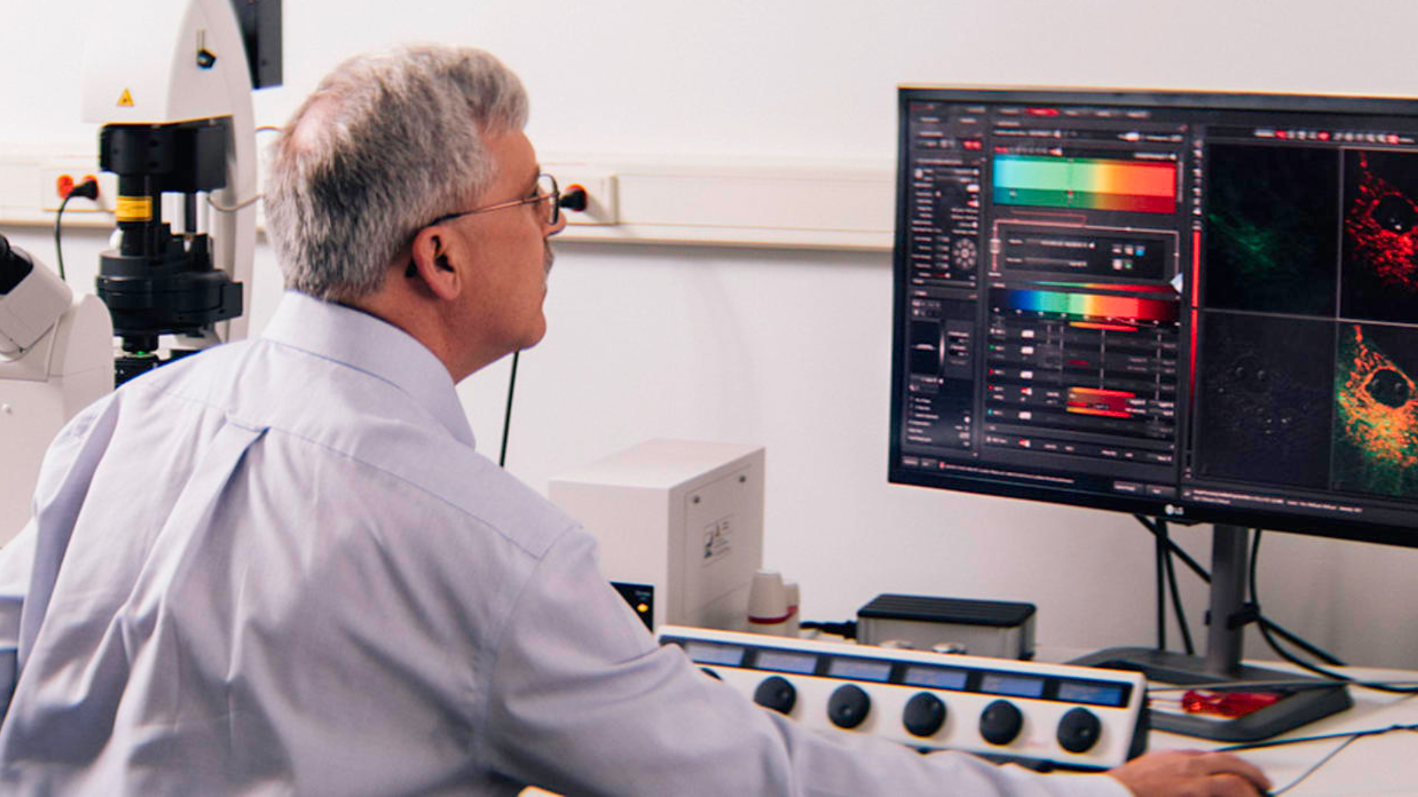
The imaging of complex cellular structures is used to determine how the temporal and spatial organization of regulatory events within cells, tissues and organisms impacts both normal and pathological processes. The state-of-the-art Imaging Shared Resource provides access to standard and advanced optical imaging systems capable of reaching these goals and offers assistance with advanced image analysis solutions. Researchers may be trained for unassisted use of all core instrumentation, while full-service assistance by facility staff is also available for quantitative or qualitative image capture. The Facility also offers expert technical assistance with experimental design to optimize imaging results, enabling users to get more out of the imaging technology.
Services
- Brightfield and Fluorescence Widefield Microscopy
- Spectral Confocal microscopy
- 2-photon intravital microscopy
- Advanced quantitative imaging (FRET, FRAP, photobleaching, colocalization)
- 2D, 3D, multichannel, multipoint, live-cell, timelapse imaging
- Small animal, whole body luminescence, fluorescence, CT and ultrasound imaging
- Low magnification and photomacrography
- Automated whole slide and plate scanning
- Personalized training and assistance on all imaging systems
- Personalized image analysis training and services
- Assistance with microscopes within individual labs
- Traditional photographic services

Equipment & Features
- Nikon Eclipse NiE motorized, automated upright microscope
- Nikon 80i upright microscope
- Nikon E600 upright microscope
- Nikon SMZ800 and 1500 stereomicroscopes
- Nikon Eclipse TiE automated, inverted microscope with environmental chamber
- Nikon TE300 semi-automated, inverted microscope with environmental chamber
- Nikon TE2000 inverted microscope
- Leica TCS SP8 X WLL laser scanning confocal microscope with environmental chamber
- Leica TCS SP5 II laser scanning confocal microscope with environmental chamber
- Leica TCS SP8 MP 2-photon intravital microscope
- Perkin Elmer IVIS SpectrumCT small animal, whole body imaging system
- Perkin Elmer / Sonovol pre-clinical ultrasound imager
- Traditional digital camera system (Nikon Z6II)
- Advanced image analysis workstations
Pricing
For pricing information, visit iLab or contact the managing director.
This facility is supported in part by a Cancer Center Support Grant (CCSG) awarded by the National Cancer Institute (NCI) to the Ellen and Ronald Caplan Cancer Center.
National Institutes of Health (NIH) has introduced new standards for grand applications to address an increasing trend of reports of failure to replicate important basic/preclinical studies.
The Wistar Institute
Imaging Facility, Room 287
3601 Spruce Street
Philadelphia, PA 19104
215-898-3887
Qing Chen, M.D., Ph.D.
Scientific Director
James Hayden, B.A., R.B.P., F.B.C.A.
Managing Director
Frederick Keeney, B.S.
Assistant Managing Director
Marie Stoltz, B.S., M.S.
Research Assistant
Monday-Friday
8:30 am-5:00 pm

