
Author: The Wistar Institute

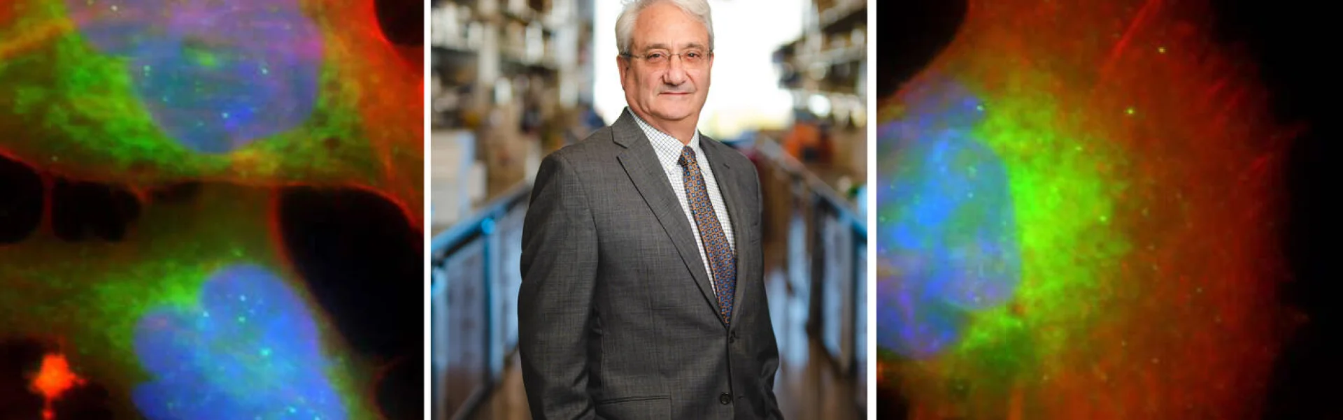
Joseph Salvino, Ph.D.

  • Professor, Molecular and Cellular Oncogenesis Program, Ellen and Ronald Caplan Cancer Center

  • Scientific Director, Molecular Screening & Protein Expression Facility

Salvino is the first medicinal chemist to join Wistar’s staff, and his work to identify novel small molecule lead compounds provides a strong basis for collaboration with all Wistar researchers.

Salvino has more than 30 years of experience in drug discovery, including early stage hit-to-lead, lead optimization, and preclinical development where several drug candidates have successfully completed human clinical trials, including Entereg®, ADL-101, and Radezolid®. Prior to joining Wistar, Salvino was a professor in the Department of Pharmacology and Physiology at Drexel University College of Medicine. Before that, he held high level positions including vice president, senior director, and director in the biotechnology and pharmaceutical industries of Sterling Winthrop, Rhone-Poulenc Rorer, Aventis, Rib-X Pharmaceuticals, Adolor Corporation, and Cephalon.

Salvino received his Ph.D. in organic chemistry from Brown University and completed postdoctoral training in synthetic and medicinal chemistry at the University of Pennsylvania under the mentorship of K.C. Nicolaou and Ralph F. Hirschmann.

View Publications

The Salvino Laboratory

215-495-6866

jsalvino@wistar.org

The Salvino Laboratory

Salvino’s research is directed at drug discovery and development. His laboratory focuses on early drug discovery and small molecule tool compounds for in vivo target validation to confirm the pharmacological relevance and “drugability” of a therapeutic target. His lab’s work in medicinal chemistry optimization is focused on the design and creation of new chemical analogs to understand structure activity relationships (SAR) critical for optimizing potency and in vivo efficacy for a lead series. This effort is important to help identify a potential preclinical drug candidate or future therapeutic agent against a new biological target.

Salvino is the Scientific Director of the Molecular Screening & Protein Expression Shared Resource which closely aligns screening capabilities for lead optimization and hit-to-lead activities that are ongoing in his laboratory.

Current projects include compounds blocking HIV and other antivirals, glutamate transporter (EAAT2) stimulators, ACSS2 Inhibitors, Proteolysis Targeting Chimeras (PROTACs) with a focus on CDK6, Mdm2, and Tert degraders, small molecules targeting protein-protein interactions, chemical probes for assay development, and cancer targeting and gram-negative bacteria targeting pro-drugs.

Staff
  • Postdoctoral Fellows

    Adi Narayana Reddy Poli, Ph.D.
    Jitendra Gour, Ph.D.
    Irfan Khan, Ph.D.
    Nitesh Nandwana, Ph.D..

  • UPenn Capstone Masters in Chemistry Students

    Heyang Shen
    Honey Shah

Salvino Lab in the News

Selected Publications

  • Targeting the CDK6 Dependence of Ph+ Acute Lymphoblastic Leukemia.

    Porazzi, P., Dominici, M.D., Salvino, J., Calabretta, B. “Targeting the CDK6 Dependence of Ph+ Acute Lymphoblastic Leukemia.” Genes (Basel). 2021 Aug 29;12(9):1355. doi: 10.3390/genes12091355.

  • Metabolic Control of Brisc-shmt2 Assembly Regulates Immune Signalling.

    Walden, M., Tian, L., Ross, R.L., Sykora, U.M., Byrne, D.P., Hesketh, E.L., Masandi, S.K., Cassel, J., George, R., Ault, J.R., Oualid, F.E., et al. “Metabolic Control of Brisc-shmt2 Assembly Regulates Immune Signalling.” Nature. 2019 Jun;570(7760):194-199. doi: 10.1038/s41586-019-1232-1. Epub 2019 May 29.

  • Targeting ACSS2 with a Transition-State Mimetic Inhibits Triple-Negative Breast Cancer Growth.

    Miller, K.D., Pniewski, K., Perry, C.E., Papp, S.B., Shaffer, J.D., Velasco-Silva, J.N., Casciano, J.C., Aramburu, T.M., Srikanth, Y.V.V., Cassel, J., et al. “Targeting ACSS2 with a Transition-State Mimetic Inhibits Triple-Negative Breast Cancer Growth.” Cancer Res. 2021 Mar 1;81(5):1252-1264. doi: 10.1158/0008-5472.CAN-20-1847. Epub 2021 Jan 7.

  • Selective inhibition of Ph-positive ALL cell growth through kinase-dependent and -independent effects by CDK6-specific PROTACs.

    Dominici, M.D., Porazzi, P., Xiao, Y., Chao, A., Tang, H., Kumar, G., Fortina, P., Spinelli, O., Rambaldi, A., Peterson, L.F., et al. “Selective inhibition of Ph-positive ALL cell growth through kinase-dependent and -independent effects by CDK6-specific PROTACs.”  Blood. 2020 Apr 30;135(18):1560-1573. doi: 10.1182/blood.2019003604.

  • Development of a Novel Inducer for Ebv Lytic Therapy.

    Tikhmyanova, N., Paparoidamis, N., Romero-Masters, J., Feng, X., Mohammed, F.S., Narayana Reddy, P.A., Kenney, S.C., Lieberman, P.M., Salvino, J.M. “Development of a Novel Inducer for Ebv Lytic Therapy.” Bioorg Med Chem Lett. 2019 Aug 15;29(16):2259-2264. doi: 10.1016/j.bmcl.2019.06.034. Epub 2019 Jun 22.

View Additional Publications

Continue reading

Ami Patel, Ph.D.

Assistant Professor, Vaccine & Immunotherapy Center

Patel researches next generation solutions for emerging infectious diseases, including DNA vaccines and DNA-encoded monoclonal antibodies.

Patel holds a B.Sc. in microbiology & immunology from McGill University, Canada, an M.Sc. in Medical Microbiology from London School of Hygiene & Tropical Medicine, University of London, U.K., and a Ph.D. in medical microbiology from the University of Manitoba, Canada. She received postdoctoral training at the San Raffaele Telethon Institute for Gene Therapy, Milan, Italy, the University of Pennsylvania and The Wistar Institute. She was promoted to research assistant professor at The Wistar Institute Vaccine & Immunotherapy Center; named a Caspar Wistar Fellow in 2020; and promoted to Assistant Professor in 2023.

View Publications

The Patel Laboratory

215-495-6813

The Patel Laboratory

The Patel laboratory focuses on vaccine and immunotherapy development against emerging infectious diseases, including study of immune mechanisms that contribute to protection from pathogens. Research in the lab employs non-viral DNA vectors for vaccine and antibody delivery. Patel’s most recent studies include DNA vaccine development against Ebola virus, Middle East respiratory syndrome coronavirus (MERS-CoV), and severe acute respiratory syndrome coronavirus 2 (SARS-CoV-2), and multiple studies in the rapidly advancing field of DNA-encoded monoclonal antibodies (DMAbs). Her preclinical animal studies have led to the successful clinical translation of anti-Ebola virus GP DNA vaccine and anti-Zika virus DMAb candidates (NCT03831503). Patel is part of a team at the Wistar Vaccine & Immunotherapy Center leading preclinical immunological studies of a SARS-CoV-2/COVID-19 DNA vaccine candidate. This vaccine candidate entered the clinic in about 10 weeks from vaccine design to FDA approval and trial initiation (NCT04336410 and NCT04447781).

Available Positions

A postdoctoral fellow position is available in the Patel lab, motivated candidates are encouraged to contact Dr. Patel at apatel@wistar.org.

Research

DNA VACCINE DEVELOPMENT AGAINST EMERGING INFECTIOUS DISEASES

Patel’s most recent studies include DNA vaccine development against Ebola virus, Middle East respiratory syndrome coronavirus (MERS-CoV), and severe acute respiratory syndrome coronavirus 2 (SARS-CoV-2), the virus that causes COVID-19 disease. Emerging respiratory pathogens including influenza A/B viruses and seasonal coronaviruses, as well as antimicrobial-resistant bacteria are also of interest. In addition to vaccine development, the lab studies potential correlates of protection (antibodies and T cells) in non-human primate samples to improve vaccine development and efficacy and bridge results from animal studies with human clinical data.

DNA-ENCODED MONOCLONAL ANTIBODY (DMAB) DELIVERY AGAINST INFECTIOUS DISEASES

Nucleic acid gene-encoded antibodies are a rapidly advancing field. Patel’s preclinical animal studies have led to the successful clinical translation of an anti-Zika virus DMAb candidate (NCT03831503). The lab’s research interests include approaches to improve DMAb protective efficacy through sequence engineering and strategies for effector function/immune cell recruitment. This includes understanding ways to modulate the early innate immune responses against DNA vectors to improve long-term duration in vivo. These studies could have interesting implications for both infectious diseases antibody delivery as well as different gene therapy approaches.

Patel Lab in the News

Selected Publications

  • Immunogenicity of a DNA vaccine candidate for COVID-19.

    Smith ,T.R.F.*, Patel, A.*, Ramos, S.*, Elwood, D., Zhu, X., Yan, J., Gary, E.N., Walker, S.N., Schultheis, K., Purwar, M., Xu, Z., Walters, J., Bhojnagarwala, P., Yang, M., Chokkalingam, N., Pezzoli, P., Parzych, E., Reuschel, E.L., Doan, A., Tursi, N., Vasquez, M., Choi, J., Tello-Ruiz, E., Maricic, I., Bah, M.A., Wu, Y., Amante, D., Park, D.H., Dia, Y., Ali, A.R., Zaidi, F.I., Generotti, A., Kim, K.Y., Herring, T.A., Reeder, S., Andrade, V.M., Buttigieg, K., Zhao, G., Wu, J.M., Li, D., Bao, L., Liu, J., Deng, W., Qin, C., Brown, A.S., Khoshnejad, M., Wang, N., Chu, J., Wrapp, D., McLellan, J.S., Muthumani, K., Wang, B., Carroll, M.W., Kim, J.J., Boyer, J., Kulp, D.W., Humeau, L., Weiner, D.B., Broderick, K.E. “Immunogenicity of a DNA vaccine candidate for COVID-19.” Nat Commun. 2020;11(1):2601. Epub 2020/05/21. doi: 10.1038/s41467-020-16505-0. PubMed PMID: 32433465.

  • Intradermal SynCon(R) Ebola GP DNA Vaccine Is Temperature Stable and Safely Demonstrates Cellular and Humoral Immunogenicity Advantages in Healthy Volunteers.

    Tebas, P., Kraynyak, K.A., Patel, A., Maslow, J.N., Morrow, M.P., Sylvester, A.J., Knoblock, D., Gillespie, E., Amante, D., Racine, T., McMullan, T., Jeong, M., Roberts, C.C., Park, Y.K., Boyer, J., Broderick, K.E., Kobinger, G.P., Bagarazzi, M., Weiner, D.B., Sardesai, N.Y., White, S.M. “Intradermal SynCon(R) Ebola GP DNA Vaccine Is Temperature Stable and Safely Demonstrates Cellular and Humoral Immunogenicity Advantages in Healthy Volunteers.” J Infect Dis. 2019;220(3):400-10. Epub 2019/03/21. doi: 10.1093/infdis/jiz132. PubMed PMID: 30891607.

  • Protective Efficacy and Long-Term Immunogenicity in Cynomolgus Macaques by Ebola Virus Glycoprotein Synthetic DNA Vaccines.

    Patel, A., Reuschel, E.L., Kraynyak, K.A., Racine, T., Park, D.H., Scott, V.L., Audet, J., Amante, D., Wise, M.C., Keaton, A.A., Wong, G., Villarreal, D.O., Walters, J., Muthumani, K., Shedlock, D.J., de La Vega, M.A., Plyler, R., Boyer, J., Broderick, K.E., Yan, J., Khan, A.S., Jones, S., Bello, A., Soule, G., Tran, K.N., He, S., Tierney, K., Qiu, X., Kobinger, G.P., Sardesai, N.Y., Weiner, D.B. “Protective Efficacy and Long-Term Immunogenicity in Cynomolgus Macaques by Ebola Virus Glycoprotein Synthetic DNA Vaccines.” J Infect Dis. 2019;219(4):544-55. Epub 2018/10/12. doi: 10.1093/infdis/jiy537. PubMed PMID: 30304515.

  • In Vivo Delivery of a DNA-Encoded Monoclonal Antibody Protects Non-human Primates against Zika Virus.

    Esquivel, R.N.*, Patel, A.*, Kudchodkar, S.B., Park, D.H., Stettler, K., Beltramello, M., Allen, J.W., Mendoza, J., Ramos, S., Choi, H., Borole, P., Asija, K., Bah, M., Shaheen, S., Chen, J., Yan, J., Durham, A.C., Smith, T.R.F., Broderick, K., Guibinga, G., Muthumani, K., Corti, D., Humeau, L., Weiner, D.B. “In Vivo Delivery of a DNA-Encoded Monoclonal Antibody Protects Non-human Primates against Zika Virus.” Mol Ther. 2019;27(5):974-85. Epub 2019/04/10. doi: 10.1016/j.ymthe.2019.03.005. PubMed PMID: 30962164; PMCIDPMC6520333.

  • In Vivo Delivery of Synthetic Human DNA-Encoded Monoclonal Antibodies Protect against Ebolavirus Infection in a Mouse Model.

    Patel, A., Park, D.H., Davis, C.W., Smith, T.R.F., Leung, A., Tierney, K., Bryan, A., Davidson, E., Yu, X., Racine, T., Reed, C., Gorman, M.E., Wise, M.C., Elliott, S.T.C., Esquivel, R., Yan, J., Chen, J., Muthumani, K., Doranz, B.J., Saphire, E.O., Crowe, J.E., Broderick, K.E., Kobinger, G.P., He, S., Qiu, X., Kobasa, D., Humeau, L., Sardesai, N.Y., Ahmed, R., Weiner, D.B. “In Vivo Delivery of Synthetic Human DNA-Encoded Monoclonal Antibodies Protect against Ebolavirus Infection in a Mouse Model.” Cell Rep. 2018;25(7):1982-93 e4. Epub 2018/11/15. doi: 10.1016/j.celrep.2018.10.062. PubMed PMID: 30428362; PMCIDPMC6319964.

View Additional Publications

Continue reading

Yulia Nefedova, M.D., Ph.D.

Associate Professor, Immunology, Microenvironment and Metastasis Program, Ellen and Ronald Caplan Cancer Center

Nefedova’s research focuses on understanding molecular mechanisms by which the bone marrow microenvironment promotes tumor survival and progression.

Nefedova obtained her M.D. and Ph.D. degrees at the Pavlov State Medical University of St. Petersburg, Russia. She then pursued postdoctoral training at the H. Lee Moffitt Cancer Center and Research Institute in Tampa, FL, where she began her research in multiple myeloma. In 2013, Nefedova joined The Wistar Institute as an assistant professor in the Tumor Microenvironment and Metastasis program, and was promoted to associate professor in the Immunology, Microenvironment and Metastasis Program in 2017.

View Publications

The Nefedova Laboratory

215-495-6952

ynefedova@wistar.org

The Nefedova Laboratory

The Nefedova laboratory focuses on understanding the role of the bone marrow microenvironment in regulating tumor progression and therapy resistance. The laboratory primarily studies multiple myeloma, a cancer of plasma cells that localizes in the bone marrow.

The laboratory is interested in mechanisms by which interactions between neutrophils in the bone marrow and multiple myeloma cells promote disease progression and chemoresistance. They are investigating the implication of neutrophil extracellular traps (NETs) — structures composed of chromatin and neutrophil proteins — in myeloma progression, and targeting a process of NET formation for treatment of multiple myeloma. The laboratory is also interested in the role of the S100A9 protein, highly expressed by neutrophils, in the pathogenesis of multiple myeloma.

Staff
  • Postdoctoral Fellow

    Cindy Lin, Ph.D.

  • Predoctoral Trainee

    Marina Li

  • Research Assistant

    Matthew Rosenwasser

Nefedova Lab in the News

Selected Publications

  • Bone marrow myeloid cells in regulation of multiple myeloma progression.

    Herlihy, S.E., Lin, C., Nefedova, Y. “Bone marrow myeloid cells in regulation of multiple myeloma progression.” Cancer Immunol Immunother. 2017 Aug;66(8):1007-1014. doi: 10.1007/s00262-017-1992-0. Epub 2017 Apr 4.

  • Lectin-type oxidized LDL receptor-1 distinguishes population of human polymorphonuclear myeloid-derived suppressor cells in cancer patients.

    Condamine, T., Dominguez, G.A., Youn, J.I., Kossenkov, A.V., Mony, S., Alicea-Torres, K., Tcyganov, E., Hashimoto, A., Nefedova, Y., Lin, C., et al. “Lectin-type oxidized LDL receptor-1 distinguishes population of human polymorphonuclear myeloid-derived suppressor cells in cancer patients.” Sci Immunol. 2016 Aug;1(2). pii: aaf8943. doi: 10.1126/sciimmunol.aaf8943. Epub 2016 Aug 5.

  • Agonist-Mediated Activation of STING Induces Apoptosis in Malignant B Cells.

    Tang, C.H., Zundell, J.A., Ranatunga, S., Lin, C., Nefedova, Y., Del Valle, J.R., Hu, C.C. “Agonist-Mediated Activation of STING Induces Apoptosis in Malignant B Cells.” Cancer Res. 2016 Apr 15;76(8):2137-52. doi: 10.1158/0008-5472.CAN-15-1885. Epub 2016 Mar 7.

  • Anti-myeloma effect of pharmacological inhibition of Notch/gamma-secretase with RO4929097 is mediated by modulation of tumor microenvironment.

    Pisklakova, A., Grigson, E., Ozerova, M., Chen, F., Sullivan, D.M., Nefedova, Y., et al. “Anti-myeloma effect of pharmacological inhibition of Notch/gamma-secretase with RO4929097 is mediated by modulation of tumor microenvironment.” Cancer Biol Ther. 2016 May 3;17(5):477-85. doi: 10.1080/15384047.2016.1156261. Epub 2016 Mar 2.

  • Bone marrow PMN-MDSCs and neutrophils are functionally similar in protection of multiple myeloma from chemotherapy.

    Ramachandran, I.R., Condamine, T., Lin, C., Herlihy, S.E., Garfall, A., Vogl, D.T., Gabrilovich, D.I., Nefedova, Y., et al. “Bone marrow PMNMDSCs and neutrophils are functionally similar in protection of multiple myeloma from chemotherapy.” Cancer Lett. 2016 Feb 1;371(1):117-24. doi: 10.1016/j.canlet.2015.10.040. Epub 2015 Nov 27.

View Additional Publications

Continue reading

Qin Liu, M.D., Ph.D.

Professor, Molecular and Cellular Oncogenesis Program, Ellen and Ronald Caplan Cancer Center

Liu applies biostatistics, the statistical analysis of complex data generated through modern biological approaches and clinical information, to find correlations between biomarkers, disease and individual patient health.

Liu earned a medical degree and a master’s degree in health statistics at Shanxi Medical University in Taiyuan, China. She obtained a Ph.D. in biostatistics at Shanghai Medical University in Shanghai, China in 1998. She then completed a postdoctoral fellowship in biostatistics and epidemiology at the University of Massachusetts. While there, she earned a second master’s degree in public health and epidemiology. In 2005, Liu was appointed assistant professor in the Department of Cancer Biology at the University of Massachusetts Medical School (UMMS). Two years later, she joined the Biostatistical Research Group in the Division of Preventive and Behavioral Medicine and served as an assistant professor of Biostatistics in the Department of Medicine at UMMS. Liu joined The Wistar Institute in 2011 as an associate professor and was promoted to professor in 2017.

View Publications

The Liu Laboratory

215-495-6940

qliu@wistar.org

The Liu Laboratory

The Liu laboratory applies biostatistics to several areas of research, including: basic cancer research and clinical trials of cancer immunotherapy; infectious diseases; behavioral and educational intervention research; and research on health care outcomes.

Staff
  • Scientific Programmer/Analyst

    Xiangfan Yin, M.S.

  • Statistical Programmer/Analyst

    Jianyi Ding, M.S.

Research

The Biostatistics Unit supervised by Liu provides in-house biostatistics expertise to accommodate continuing growth in translational and pre-clinical cancer research. This Unit provides statistical support to biological laboratories at Wistar and its tasks include, but not limited to, large data management, experiment design, statistical data analysis, data presentation for manuscripts and grant proposal development. Liu has extensive experience working with basic and clinical investigators on statistical issues. Her strong statistical and biomedical backgrounds have enabled her to provide exceptionally high levels of scientific input to different projects. As a result, her effort has contributed to many published papers and funded grant proposals for clinical and basic science investigators. Within a short time after she joined The Wistar Institute, she engaged in a plethora of collaborative projects with Wistar Cancer Center members and obtained joint NIH funding with inside and outside Wistar investigators, including two P01 Program Project grants on novel molecular therapies of prostate cancer and melanoma targeted therapies. There is significant institutional support for collaborative efforts between the Biostatistics Unit and the investigators at Wistar.

Selected Publications

  • Evaluation of drug combination effect using a Bliss independence dose-response surface model.

    Liu, Q., Yin, X., Languino, L.R., Altieri, D.C. “Evaluation of drug combination effect using a Bliss independence dose-response surface model.” Statistics in Biopharmaceutical Research 2018.10:2, 112-122, DOI: 10.1080/19466315.2018.1437071.

  • Hepatitis C virus modulates IgG glycosylation in HIV co-infected antiretroviral therapy suppressed individuals.

    Giron, L.B., Azzoni, L., Yin, X., Lynn, K.M., Ross, B.N., Fair, M., Damra, M., Sciorillo, A.C., Liu, Q., Jacobson, J.M., Mounzer, K., Kostman, J.R., Abdel-Mohsen, M., Montaner, L.J., Papasavvas, E. “Hepatitis C virus modulates IgG glycosylation in HIV co-infected antiretroviral therapy suppressed individuals.” AIDS. 34(10): 1461-1466, Jul 2020.

  • The mitophagy effector FUNDC1 controls mitochondrial reprogramming and cellular plasticity in cancer cells.

    Li, J., Agarwal, E., Bertolini, I., Seo, J.H., Caino, M.C., Ghosh, J.C., Kossenkov, A.V., Liu, Q., Tang, X-Y, Goldman, A.R., Languino, L.R., Speicher, D.W., Altieri, D.C. “The mitophagy effector FUNDC1 controls mitochondrial reprogramming and cellular plasticity in cancer cells.” Science Signaling. 2020; 13 (642), e aaz8240, DOI: 10.1126/scisignal.aaz8240.

  • The αvβ6 integrin in cancer cell-derived small extracellular vesicles enhances angiogenesis.

    Krishn, S.R., Salem, I., Quaglia, F., Naranjo, N., Agarwal, E., Liu, Q., Sarker, S., Kopenhaver, J., McCue, P.A., Weinreb, P.H., Violette, S.M., Altieri, D.C., Languino, L.R. “The αvβ6 integrin in cancer cell-derived small extracellular vesicles enhances angiogenesis.” Journal of extracellular vesicles. 2020; 9:1, 1763594, DOI: 10.1080/20013078.2020.1763594

  • African-centric variant in TP53 is associated with increased iron accumulation and bacterial pathogenesis but improved response to malaria toxin.

    Singh, K.S., Leu, J.I., Barnoud, T., Vonteddu, P., Gnanapradeepan, K., Lin, C., Liu, Q., Barton, J.C., Kossenkov, A.V., George, D.L., Murphy, M.E., Dotiwala, F. “African-centric variant in TP53 is associated with increased iron accumulation and bacterial pathogenesis but improved response to malaria toxin.” Nature Communications. 2020 Jan 24;11(1):473. doi: 10.1038/s41467-019-14151-9. PMID: 31980600. PMCIDPMC6981190.

View Additional Publications

Continue reading

Daniel Kulp, Ph.D.

Associate Professor, Vaccine & Immunotherapy Center

Kulp has more than 15 years of experience developing molecular design software and leading protein engineering projects. He joined Wistar from The Scripps Research Institute and International AIDS Vaccine Initiative where he was a principal scientist.

Kulp received a bachelor’s degree in Computer Science and Molecular Biology & Biochemistry from Rutgers, The State University of New Jersey, followed by a Ph.D. in Biochemistry and Molecular Biophysics from the University of Pennsylvania. He completed postdoctoral training in structure-based and experimental protein engineering at Los Alamos National Laboratory.

View Publications

The Kulp Laboratory

215-898-6587

dwkulp@wistar.org

The Kulp Laboratory

The Kulp laboratory focuses on rational vaccine and therapeutic antibody design for a variety of priority infectious diseases (e.g. Lassa Virus, HIV, Influenza) and cancer targets. The ultimate test of the lab’s understanding of B cell immune responses is to design new immunogens that drive predictable antibody maturation. To that end, the lab is interested in the development and application of protein engineering methods for modifying antigen/cell receptor interfaces, antigen/antibody interfaces, antigen surface properties and core stabilization.

Staff
  • Postdoctoral Fellow

    Jinwei Huang

  • Ph.D. Graduate Students

    Michaela Helble
    Kylie Konrath
    Rumi Habib
    Niklas Laegner
    Shahlo Solieva
    Yuanhan Wu

  • M.S. Graduate Student

    Sarah Kim

  • Research Assistants

    Kelly Bayruns
    Amber Kim
    Joyce Park
    Madison McCanna

  • Research Apprentice

    Alex Dalton


Lab Alumni

Susanne Walker – Merck
Neethu Chokkalingkam – Ocugen
Sinja Kriete – Lake Erie College of Osteopathic Medicine (D.O. Student)
Nicholas Shupin – Robert Wood Johnson Medical School (M.D. student)
Alana Hyunh – University of Rochester (Ph.D. student)

Kulp Lab in the News

  • Wistar Scientists Successfully Engineer a Goldilocks Construct: Therapeutic Antibody Could Be a Future Medicine to Improve Outcomes for Melanoma

    Featured News

  • Highlighting Vaccine Research at The Wistar Institute Through the Penn-CHOP-Wistar Vaccine Symposium

    Featured News

  • Novel Nanoparticle SARS-CoV-2 Vaccine Combines Immune Focusing and Self-assembling Nanoparticles to Elicit More Potent Protection

    Press Release

Selected Publications

View Additional Publications

Continue reading

Andrew Kossenkov, Ph.D.

  • Assistant Professor, Vaccine & Immunotherapy Center

  • Gene Expression and Regulation Program, Ellen and Ronald Caplan Cancer Center

  • Scientific Director, Bioinformatics Facility 

Kossenkov applies computational approaches to the analysis of various kinds of biomedical high-throughput data in effort to interpret results and visualize complex data.

He obtained his bachelor’s and master’s degrees in computer science and bioinformatics from Moscow Engineering Physics Institute in Russia, and his Ph.D. in biomedical science from Drexel University in Philadelphia. In 2007, Kossenkov joined The Wistar Institute as a postdoctoral fellow in the Showe lab and later became managing director of the Bioinformatics Facility. He was appointed assistant professor in 2019.

View Publications

The Kossenkov Laboratory

215-495-6898

akossenkov@wistar.org

The Kossenkov Laboratory

The Kossenkov laboratory collaborates with several other Wistar labs on research projects in cancer, infectious diseases and basic biological questions, including regeneration and stem cell biology, mitochondrial proteins, transcriptional regulation, synapse biology, blood development, and global epigenetic gene silencing. The lab uses bioinformatics, biostatistics and computational biology approaches in a variety of models. They rely heavily on bioinformatics analysis of drug screen results, next-generation sequencing data and proteomics.

Research

Through a network of internal and external collaborations, the Kossenkov lab provides bioinformatics expertise for high throughput data support, analysis and annotation and complex results visualization. Thanks to Kossenkov’s experience in development of database-oriented gene annotation pipelines and algorithms to visualize highly specific and intricate results, the lab provides flexible and customizable support for a wide range of experiments.

Research collaborations outside of Wistar include teams at Fox Chase Cancer Center, The Children’s Hospital of Philadelphia, the University of Pennsylvania, Drexel University, New York University, Weill Cornell, Harvard, and others.

Kossenkov Lab in the News

Selected Publications

  • A Gene Expression Classifier from Whole Blood Distinguishes Benign from Malignant Lung Nodules Detected by Low-Dose CT

    Kossenkov, A.V., Qureshi, R., Showe, L.C., et al. “A Gene Expression Classifier from Whole Blood Distinguishes Benign from Malignant Lung Nodules Detected by Low-Dose CT”. Cancer Res. 2019 Jan 1;79(1):263-273. doi: 10.1158/0008-5472.CAN-18-2032. Epub 2018 Nov 28.

  • Unique pattern of neutrophil migration and function during tumor progression.

    Patel, S., Fu, S., Mastio,  J., Dominguez, G.A., Purohit, A., Kossenkov, A., Lin, C., Alicea-Torres, K., Sehgal, M., Nefedova, Y., Zhou, J., Languino, L.R., Clendenin, C., Vonderheide, R.H., Mulligan, C., Nam, B., Hockstein, N., Masters, G., Guarino, M., Schug, Z.T., Altieri, D.C., Gabrilovich, D.I. “Unique pattern of neutrophil migration and function during tumor progression.” Nat Immunol. 2018 Nov;19(11):1236-1247. doi: 10.1038/s41590-018-0229-5. Epub 2018 Oct 15

  • IRE1α-XBP1 controls T cell function in ovarian cancer by regulating mitochondrial activity.

    Song, M., Sandoval, T.A., Chae, C.S., Chopra, S., Tan, C., Rutkowski, M.R., Raundhal, M., Chaurio, R.A., Payne, K.K., Konrad, C., Bettigole, S.E., Shin, H.R., Crowley, M.J.P., Cerliani, J.P., Kossenkov, A.V., Motorykin, I., Zhang, S., Manfredi, G., Zamarin, D., Holcomb, K., Rodriguez, P.C., Rabinovich, G.A., Conejo-Garcia, J.R., Glimcher, L.H., Cubillos-Ruiz, J.R. “IRE1α-XBP1 controls T cell function in ovarian cancer by regulating mitochondrial activity.” Nature. 2018 Oct;562(7727):423-428. doi: 10.1038/s41586-018-0597-x. Epub 2018 Oct 10.

  • CARM1-expressing ovarian cancer depends on the histone methyltransferase EZH2 activity.

    Karakashev, S., Zhu, H., Wu, S., Yokoyama, Y., Bitler, B.G., Park, P.H., Lee, J.H., Kossenkov, A.V., Gaonkar, K.S., Yan, H., Drapkin, R., Conejo-Garcia, J.R., Speicher, D.W., Ordog, T., Zhang, R. “CARM1-expressing ovarian cancer depends on the histone methyltransferase EZH2 activity.” Nat Commun. 2018 Feb 12;9(1):631. doi: 10.1038/s41467-018-03031-3.

  • RNA-Seq of Kaposi’s sarcoma reveals alterations in glucose and lipid metabolism.

    Tso, F.Y., Kossenkov, A.V., Wood, C., et al. “RNA-Seq of Kaposi’s sarcoma reveals alterations in glucose and lipid metabolism.” PLoS Pathog. 2018 Jan 19;14(1):e1006844. doi: 10.1371/journal.ppat.1006844. eCollection 2018 Jan.

View Additional Publications

Continue reading

With Support from the National Science Foundation, Wistar Launches its First National, College-level Biomedical Research Internship Program

Inaugural class learns how to conduct research in immersive summer program

If you take a walk along any one of Wistar’s corridors in the summer, chances are you will see students at the laboratory bench working intently on their research projects. This summer is no exception and includes a select group of college students arriving in Philadelphia for a 12-week Wistar summer intensive research experience.

In the new Research Experiences for Undergraduates (REU) Program students are fully immersed in biomedical science experiments under the guidance of a Wistar mentor-scientist leading one of 33+ active labs at the Institute. Students conduct innovative research, applying state-of-the-science techniques to plan and execute experiments and advance their projects. REU is funded by the National Science Foundation (NSF), organized to encourage STEM student innovators to pursue graduate education and eventual careers that will advance the life sciences into new directions.

Of the inaugural group of 16 students, 12 identified as female and five identified as male. 10 students were funded by the NSF REU grant and six were funded through a PAsmart grant. All NSF REU students were from underrepresented groups and participating from California, Delaware, Florida, Maryland, Massachusetts, New Jersey, Pennsylvania, and Texas.

“Every day Wistar invests in pioneering research accomplished by our groundbreaking scientists. That investment includes training our next-gen scientists. These students will become leading scientists in the future and will be our answer and our success. Their discoveries will become tomorrow’s life-saving therapies,” said Dr. Kristy Shuda McGuire.

Since joining Wistar, dean of Biomedical Studies Dr. Shuda McGuire’s singular focus has been to expand education and training programs to impact as many as possible underserved and underrepresented students from the surrounding greater Philadelphia community and now, with the support of the NSF from around the country.

Inaugural REU students Rickelle Wescott and Kiara Garcia Castro shared their experiences in the REU Program where days are filled learning biomedical research lab methods. Nights are for relaxing (in communal housing provided through the Program) and reading science articles, deep dives into more research, and nailing down protocols for the next day’s experiments. Eat, sleep and repeat.

Originally from Puerto Rico, Kiara always aspired to a science job. She’s a rising junior biology major at Temple University. She knew she wanted to do research but did not know where to start.

“Wistar is training friendly and accommodating to everyone. My mentor emphasized it was okay to make mistakes and not feel bad when you mess up because you are learning how to fix your mistakes. He emphasized all researchers have gone through this—you aren’t born knowing how to do research,” said Kiara.

Their diligence in the Program pays off as these next-gen scientists push past their fears about undertaking hugely complex science.

“What’s been the most challenging was to understand and absorb all this scientific information in a short amount of time and then to apply it. I had to understand the purpose of everything: the different solutions, different terms and experiments. That was the difficult part. You just try to be quick on your feet,” said Rickelle.

Rickelle, who is on the pre-med track at Hampton University, is undertaking a variety of research experiments far different from anything she’s done so far in her college science curriculum.

“At Wistar, you feel part of something that is making breakthroughs. You feel like you can be someone who can make a discovery too,” said Rickelle.

Genomic Origami: Wistar Scientist Dr. Kavitha Sarma Studies How the Shape of Our Genes Impacts Disease

A Q&A with Dr. Kavitha Sarma

Dr. Kavitha Sarma runs an independent lab focused on nucleic acid structures called R-loops that contain both DNA and RNA and assist in gene expression. Dr. Sarma, associate professor in Wistar’s Gene Expression and Regulation Program, recently published a paper in Molecular Cell about genomic structures — specifically, G-quadruplexes and R-loops.

R-loops are bubble-like structures that can form in our DNA, and they can affect how genes are expressed — whether genes are turned on or off. G-quadruplexes form on the single-strand DNA of R-loops and can stabilize R-loops. In her research, Dr. Sarma found that R-loops and G-quadruplexes can influence the binding of a protein called CTCF, which helps fold and organize our DNA. This folding process is important for gene expression. If the genome is folded correctly, that allows genes to be expressed the way they should be. But if the genome is folded incorrectly, it can cause faulty patterns of gene expression, which can potentially lead to disease and cancer. R-loops and G-quadruplexes can play a role in cancer and disease by recruiting CTCF in a way that promotes faulty gene expression.

IN YOUR PAPER, YOU FOUND THAT R-LOOPS AND G4S HAD AN INTERESTING RELATIONSHIP WITH A CERTAIN MOLECULE. COULD YOU EXPLAIN THAT FINDING?

In every cell nucleus in your body, you have something like two meters of DNA, if you were to unravel it completely into one long double helix. Just to make genetic information physically fit in your body, the genome has to be compacted, and that needs to happen in every single cell, too.

There are many proteins that function in genome folding. We found that R-loops and G4s can influence the binding of one of these proteins – CTCF, which has a very important role in how the genome is folded.

This folding process, which also serves as a kind of information organization process, is important for how cells develop and specialize in our body. For example, the way a neuron’s genome is folded and expressed will be different from the genome folding and gene expression of a pancreatic cell because the two cell types fulfill different purposes. Epigenetic regulation from factors like genome folding allows for a diversity of gene expression — which, in turn, allows for a diversity of cell types and functions.

So, if a genome is folded correctly in the nucleus and the right regions are next to each other, that has a positive effect, and genes are expressed the way they should be. But if CTCF folds the genome incorrectly — for example, if R-loops and G4s form and facilitate CTCF binding to regions where it isn’t supposed to bind — we might see incorrect patterns of gene expression and the kinds of dysregulation you’d find in cancer and disease.

WHAT ARE THE PATHOGENIC IMPLICATIONS OF CTCF RECRUITMENT?

This finding, that G4s affect CTCF, tells us that the genome misfolding in disease can be at least partially due to the formation of R-loop structures. In addition to developmental disorders, R-loop and G4 structures can play problematic roles in cancer because they’re what we call co-transcriptional. When transcription happens, these structures tend to accumulate — they tend to become stabilized. Hypertranscription that occurs in many cancers can contribute to genome misfolding through R-loop and G4 formation, which can further reinforce faulty gene expression patterns by essentially rewiring the genome.

WHAT DOES THIS REWIRING CYCLE TELL US ABOUT THE EPIGENETICS OF CANCER AND DISEASE?

I think that this research gives us a good roadmap for looking for therapeutics down the line, because a better understanding of epigenetic regulation gives us deeper insight into how disease states work at a very localized level.

We know that R-loops and G4s can alter CTCF binding and change genome folding. Going forward, we can identify pathogenic contacts that occur because of these genomic structures and try to correct them. This is how foundational research — understanding processes that weren’t understood before — can lead to advances in the science of human health.

Aaron R. Goldman, Ph.D.

Assistant Professor, Molecular and Cellular Oncogenesis Program, Ellen and Ronald Caplan Cancer Center

Goldman applies mass spectrometry (MS)-based approaches, primarily metabolomics and lipidomics, to examine metabolism in a wide-range of biomedical studies applied to cancer and other human diseases as well as fundamental mechanistic biology.

Goldman received his B.S. in Biological Sciences from Carnegie Mellon University. He earned a Ph.D. in Cell and Molecular Biology from Stanford University. Goldman joined The Wistar Institute in 2014 as a postdoctoral fellow in David Speicher’s laboratory where he utilized mass spectrometry-based proteomics, lipidomics, and metabolomics to study melanoma, ovarian cancer, and other cancers. He was subsequently appointed as Associate Managing Director of Metabolomics and Lipidomics in the Proteomics and Metabolomics Shared Resource. In 2022, Goldman was promoted to Assistant Professor in the Molecular and Cellular Oncogenesis Program of Wistar’s Ellen and Ronald Caplan Cancer Center.

View Publications

The Goldman Laboratory

agoldman@wistar.org

The Goldman Laboratory

The Goldman laboratory extensively utilizes state-of-the-art high resolution mass spectrometry to study metabolic and lipid changes in cancer and infectious diseases in collaboration with researchers at Wistar and many other institutions. Most projects are systems-level studies that assimilate data from multiple sources to better understand the mechanisms underlying diseases, to identify potential therapeutic targets, and to uncover putative diagnostic and prognostic biomarkers.

Metabolomics and lipidomics are the study of the set of polar metabolites and lipids (non-polar metabolites), respectively, in a biological system. Unbiased profiling of these small molecules provides insight into metabolic pathways and processes perturbed in disease states that could lead to testable hypotheses for disease progression or therapy response. Alternatively, these methods can confirm findings suggested by orthogonal assays such as transcriptomics or proteomics. Use of these techniques has exponentially increased in recent years as mass spectrometry technologies have enabled greater depth of analysis and more confident identification of specific metabolites and lipids.

Available Positions

Motivated candidates are encouraged to inquire about the positions below by contacting Dr. Goldman at agoldman@wistar.org.

A Postdoctoral Fellow and a Research Assistant position are available to work on multiple ongoing projects in the Goldman laboratory. The research focus of these positions could include (depending upon interest and expertise): small molecule omics studies of melanoma tumor progression and resistance to therapies; pregnancy biomarker validation; and/or implementation of new and improved mass spectrometry-based analyses of polar metabolites and lipids (see project descriptions above). Postdoctoral Fellow candidates should have recently received or be close to obtaining their Ph.D. degree (or equivalent) and have a strong background in small molecule mass spectrometry. Research Assistant candidates should have a B.S. degree in biochemistry or equivalent. Experience with MS is preferred but not required.

Research

The Goldman laboratory is focused on improving both mass spectrometry-based metabolomics and lipidomics methods and applying these state-of-the-art methods to a wide-range of collaborative studies investigating cancers including ovarian, pancreatic, prostate, melanoma, and leukemia, as well as infectious diseases including HIV and COVID-19. A combination of untargeted and targeted approaches both at steady-state and using isotopic labeling (flux analysis) have led to important findings associated with disease progression and treatment, including changes in energy metabolism, perturbation of specific macromolecule biosynthesis pathways, and extensive lipid remodeling. Results of mass spectrometry studies have often been used to validate findings from RNAseq and other orthogonal approaches, and integration with other data sets has yielded combinatorial biomarkers.

Available Positions

Motivated candidates are encouraged to inquire about the positions below by contacting Dr. Goldman at agoldman@wistar.org.

A Postdoctoral Fellow and a Research Assistant position are available to work on multiple ongoing projects in the Goldman laboratory. The research focus of these positions could include (depending upon interest and expertise): small molecule omics studies of melanoma tumor progression and resistance to therapies; pregnancy biomarker validation; and/or implementation of new and improved mass spectrometry-based analyses of polar metabolites and lipids (see project descriptions above). Postdoctoral Fellow candidates should have recently received or be close to obtaining their Ph.D. degree (or equivalent) and have a strong background in small molecule mass spectrometry. Research Assistant candidates should have a B.S. degree in biochemistry or equivalent. Experience with MS is preferred but not required.

Major areas of interest:

LIPID REMODELING IN TUMOR PROGRESSION AND THERAPY RESISTANCE IN MELANOMA

A primary focus of the Goldman laboratory is collaborating with other researchers to define the role that lipid metabolism plays in melanoma progression and therapy resistance. In one such academic collaboration with researchers at the University of Pennsylvania, specific lipid metabolism pathways were implicated as being affected by lysosomal autophagy inhibition by using a combination of proteomic and lipidomic approaches. Efforts are ongoing to define the mechanisms by which autophagy modulation regulates lipid metabolism using small molecule activators and inhibitors. Combinatorial inhibition of lipid metabolism pathways and lysosomal autophagy is being pursued as a putative therapeutic strategy to mitigate therapy resistance in melanoma.

THE ROLE OF THE GUT MICROBIOME IN DISEASE

There is a growing field of research on the interplay between gut microbiota and human health. Recent work has found that the gut microbiome plays roles in tumor biology, infectious diseases, and immune response. Collaborative projects at Wistar led by the laboratories of Drs. Rahul Shinde and Mohamed Abdel-Mohsen have uncovered links between gut microbiota and gut-derived products with pancreatic cancer and COVID-19, respectively. Examples of key gut-derived products uncovered by untargeted metabolomic profiling are trimethylamine N-oxide, which is produced primarily from dietary choline, and various metabolites of tryptophan. The Goldman laboratory is actively developing and implementing methods to expand coverage of gut-derived products to 1) short-chain fatty acids (SCFA; two to six carbons) that regulate cell homeostasis and have been implicated in multiple cancers and other diseases such as HIV and 2) bile acids that are involved in lipid digestion and absorption and are subject to on-going research for their roles in cancer development and progression as well as potential therapeutic applications.

DISCOVERY AND VALIDATION OF EARLY PREGNANCY BIOMARKERS

A major clinical challenge in early pregnancy is to distinguish normal intrauterine pregnancy (IUP) from abnormal conditions when ultrasound is not diagnostic. Women with early-stage pregnancy who experience abdominal pain and vaginal bleeding might have: 1) an ongoing viable IUP, 2) a non-viable intrauterine pregnancy or spontaneous abortion (SAB), or 3) ectopic pregnancy (EP). EP occurs in 1-2% of pregnant women and causes 6% of pregnancy-related deaths, while SAB affects 10-20% of pregnancies. Because clinical treatment for these possible outcomes differs greatly, it is critical that an accurate diagnosis is made as early as possible. With researchers from the University of Pennsylvania, we are performing mass spectrometry-based studies to identify putative early pregnancy protein, metabolite and/or lipid biomarkers. The end goal is to develop a novel, multiplexed, MS-based plasma biomarker test for early and accurate diagnosis of pregnancy outcome. Targeted proteomics is being used to validate previously identified candidate biomarkers with a focus on distinguishing protein isoforms to increase accuracy, and discovery metabolomics and lipidomics are being pursued to identify additional targets that may complement protein markers in biomarker panels for early pregnancy outcomes.

EMERGING MS TECHNOLOGIES FOR SMALL MOLECULE STUDIES

Ion Mobility (IM). Goldman and his team are actively evaluating different IM implementations for metabolite and lipid studies. A major challenge in the analysis of small molecules is confident annotation of isomers (molecules with the same chemical formula) and isobars (molecules with the same nominal mass at a given resolution). These compounds are indistinguishable by accurate mass, and MS/MS fragmentation is often information-poor and does not generate diagnostic fragments that are unique to a given species. They may also not be resolved by chromatographic separation. A promising emerging technology to distinguish these types of compounds is IM, which separates ions in the gas phase based on their collisional cross-section (CCS), a property derived from the mobility of the ions in an electric field. In addition to separating currently unresolved species, IM provides a fourth dimension of data to annotate detected compounds more confidently in terms of retention time, accurate mass, MS/MS spectra, and CCS. This technology is also applicable to proteomics, where it can be used to separate isomeric peptides ¬– such as those that are phosphorylated on different residues – and reduce sample complexity to increase depth of analysis.

MS Imaging. Goldman and his team are evaluating alternative instruments and approaches for tissue imaging of metabolites, lipids, and other biomolecules. Tumors are heterogenous and determination of the spatial distribution of biomolecules may provide insights into disease states and allow for stratification of patients by tumor molecular signatures for tailored treatment options. Imaging mass spectrometry (MS) uses a matrix-assisted laser desorption/ionization (MALDI) source to visualize lipids, metabolites, and peptides directly or visualize proteins and glycans with additional processing (enzymatic digestion) at 5-10 µm resolution. Analyte patterns can be compared between conditions to identify important biomolecules and define molecular signatures. These data can be used to select regions for more in-depth study using laser microdissection followed by traditional LC-MS approaches and correlated with orthogonal assays such as immunohistochemistry. Recent instruments support IM and MS/MS fragmentation to allow for higher confidence annotation of analytes.

Selected Publications

  • A Cancer Ubiquitome Landscape Identifies Metabolic Reprogramming as Target of Parkin Tumor Suppression.

    Agarwal, E., Goldman, A.R., Tang, H.Y., Kossenkov, A.V., Ghosh, J.C., Languino, L.R., Vaira, V., Speicher, D.W., Altieri, D.C. “A Cancer Ubiquitome Landscape Identifies Metabolic Reprogramming as Target of Parkin Tumor Suppression.” Sci Adv. 2021 Aug 25;7(35):eabg7287. doi: 10.1126/sciadv.abg7287. Print 2021 Aug.

  • ATF3 Coordinates Serine and Nucleotide Metabolism to Drive Cell Cycle Progression in Acute Myeloid Leukemia.

    Di Marcantonio, D., Martinez, E., Kanefsky, J.S., Huhn, J.M., Gabbasov, R., Gupta, A., Krais, J.J., Peri, S., Tan, Y., Skorski, T., et al. “ATF3 Coordinates Serine and Nucleotide Metabolism to Drive Cell Cycle Progression in Acute Myeloid Leukemia.” Mol Cell. 2021 Jul 1;81(13):2752-2764.e6.doi: 10.1016/j.molcel.2021.05.008. Epub 2021 Jun 2.

  • Plasma Markers of Disrupted Gut Permeability in Severe COVID-19 Patients.

    Giron, L.B., Dweep, H., Yin, X., Wang, H., Damra, M., Goldman AR, Gorman N, Palmer CS, Tang HY, Shaikh MW, et al. “Plasma Markers of Disrupted Gut Permeability in Severe COVID-19 Patients.” Front Immunol. 2021 Jun 9;12:686240. doi: 10.3389/fimmu.2021.686240. eCollection 2021.

  • Changes in Aged Fibroblast Lipid Metabolism Induce Age-Dependent Melanoma Cell Resistance to Targeted Therapy via the Fatty Acid Transporter FATP2.

    Alicea, G.M., Rebecca, V.W., Goldman, A.R., Fane, M.E., Douglass, S.M., Behera, R., Webster, M.R., Kugel, C.H. 3rd, Ecker, B.L., Caino, M.C., et al. “Changes in Aged Fibroblast Lipid Metabolism Induce Age-Dependent Melanoma Cell Resistance to Targeted Therapy via the Fatty Acid Transporter FATP2.” Cancer Discov. 2020 Sep;10(9):1282-1295. doi: 10.1158/2159-8290.CD-20-0329. Epub 2020 Jun 4.

  • PPT1 Promotes Tumor Growth and Is the Molecular Target of Chloroquine Derivatives in Cancer.

    Rebecca, V.W., Nicastri, M.C., Fennelly, C., Chude, C.I., Barber-Rotenberg, J.S., Ronghe, A., McAfee, Q., McLaughlin, N.P., Zhang, G., Goldman, A.R., et al. “PPT1 Promotes Tumor Growth and Is the Molecular Target of Chloroquine Derivatives in Cancer.” Cancer Discov. 2019 Feb;9(2):220-229. doi: 10.1158/2159-8290.CD-18-0706. Epub 2018 Nov 15.

View Additional Publications

Continue reading

Alessandro Gardini, Ph.D.

Associate Professor, Gene Expression and Regulation Program, Ellen and Ronald Caplan Cancer Center

Gardini studies the epigenetic control of transcription during cell differentiation and oncogenesis.

Born and raised in Italy, Gardini obtained a B.S./M.S. in medical biotechnology at the University of Bologna and attended the graduate school of Molecular Medicine at the University of Milan. He trained as a postdoctoral fellow with Dr. Ramin Shiekhattar at the Center of Genomic Regulation in Barcelona, The Wistar Institute and the University of Miami Medical School. He joined Wistar as an assistant professor in 2015. Gardini is a scholar of the Leukemia Research Foundation and the American Cancer Society.

View Publications

The Gardini Laboratory

215-898-3785

agardini@wistar.org

The Gardini Laboratory

Transcriptional regulation is a multi-layer process carried out by specialized transcription factors, co-factors and enzymatic machineries working within the context of a structured chromatin landscape.

We use a combination of genomics, proteomics and biochemistry to investigate how basal transcriptional complexes and epigenetic modulators orchestrate gene expression in normal and tumor cells.

Staff
  • Postdoctoral Fellows

    Sandra Deliard, Ph.D.

  • Graduate Students

    Luca Grillini (UniBO)
    Connor Hill (UPenn-CAMB)
    Ilan Kirkel (UniBO)
    Sarah Offley (UPenn-CAMB)
    Martina Gatto (UniBO)

  • Research Assistant

    Francis Picone


Alumni

Marco Trizzino, Ph.D. (2016-2019), assistant professor, Thomas Jefferson University

Elisa Barbieri, Ph.D. (2015-2019), Marie Sklodowska-Curie Fellow, University of Edinburgh


Available Positions

Motivated candidates are encouraged to inquire about the positions below. Contact agardini@wistar.org.

Postdoctoral Fellow
Graduate Students (BGS-UPenn)

Research

  • 1 – Enhancer Regulation During Lineage and Tissue Specification

    We dissect the transcriptional mechanisms that control fate choice and differentiation of human hematopoietic cells, particularly in the myeloid compartment. We are especially interested in the role of transcriptional enhancers.

    Enhancers are distal regulatory elements, scattered throughout the entire genome, that direct gene regulation during development. Furthermore, enhancers are active spots for transcription of long noncoding eRNAs (Gardini and Shiekhattar, 2015). These short-lived, non-polyadenylated transcripts are required to establish a link between the promoter and the distant regulatory enhancer (chromosomal looping) and contribute to the expression of the target protein-coding gene. The underlying mechanisms are poorly understood and the potential contribution of enhancers and eRNAs to cancer has just started to emerge. The lab is keen on understanding how enhancer networks get activated and promote differentiation of myeloid cells, and how this process is disrupted during leukemogenesis. We recently uncovered a novel enhancer regulatory axis in myeloid progenitor cells (Barbieri, Trizzino et al., 2018), centered around the EGR-1 transcription factor and a newly characterized module of the Integrator complex.

    Additional studies (Trizzino, Zucco et al., 2021) revealed that EGR-1 also coordinates a repressive activity, independent of Integrator, in mature macrophages. Particularly, EGR-1 restrains the activity of inflammatory enhancers and effectively curbs the response of macrophages to inflammatory stimuli.

    Download the image here.

  • 2 – Role of Protein Phosphatases in RNA Polymerase Pause-Release and Elongation

    Eukaryotic transcription pivots around the balanced activity of kinases and phosphatases that regulate the transcription initiation, early elongation and termination checkpoints. While the dynamic recruitment of kinases (i.e. CDK7, CDK9) and their cognate cyclins have been studied for many years, the association of phosphatases with active transcription has been largely understudied. We recently uncovered that protein phosphatase 2A (PP2A) is recruited at the pause-release checkpoint where it opposes CDK9 activity (Vervoort, Welsh et al., 2021). We found that the Integrator subunit INTS6 draws PP2A to the transcription bubble to dephosphorylate the RNAPII-CTD (especially Ser2) and maintain polymerase in a paused conformation. PP2A activity is frequently lost in cancer and restoring its ability to curb transcription opens up new therapeutic strategies for tumors ‘addicted’ to transcriptional elongation.

    Download the image here.

  • 3 – Integrator: The Swiss Army Knife of Transcription

    Integrator is a large, evolutionarily conserved, multiprotein complex that is quickly emerging as a centerpiece of transcriptional regulation in higher eukaryotes (Welsh and Gardini, 2022). The Integrator complex is a regulatory hub for several transcriptional and epigenetic processes. For instance, Integrator’s INTS11 subunit controls release of RNA Polymerase II at promoters via endonucleolytic cut of nascent RNA (Gardini et al., 2014Beckedorff et al., 2020). In addition, INTS11 regulates enhancer function through the processing of noncoding eRNAs (Lai, Gardini et al., 2015). We identified a new functional module of Integrator, centered around the INTS13 subunit, that prompts enhancer activation in myeloid cells (Barbieri, Trizzino et al., 2018). More recently, we showed that the INTS6/INTS8 subunits of the complex recruit the PP2A phosphatase to chromatin, to control phosphorylation of the RNAPII-CTD as well as other polymerase co-factors implicated in pausing and elongation (Vervoort, Welsh et al., 2021). Notably, Integrator subunits are found mutated in developmental diseases and are frequently lost in certain tumor types. The lab keeps pursuing the biochemical and functional dissection of all 14 subunits of the Integrator complex.

    Download the image here.

  • 4 – Profiling Human Transcriptomes at Better Resolution

    Conventional transcriptomic analyses (i.e. ribo-depleted RNA-seq, 3’-seq) offer a glimpse into global gene regulation but often fail to convey the most accurate picture. Because RNA-seq captures steady-state and cytoplasmic transcripts, changes in gene expression can be hindered by RNA stability. Furthermore, RNA Polymerase dynamics is only revealed by real-time nascent RNA transcription. We strive to implement and optimize techniques of nascent RNA-seq, improving their reproducibility and applicability. To this purpose, we recently developed fastGRO (Barbieri, Hill et al., 2020) as a global nuclear run-on assay of rapid execution and suitable for primary cells.

    Download the image here.

  • 5 – Role of ARID1A in Transcriptional Regulation

    Chromatin remodelers regulate DNA accessibility by positioning nucleosomes at active promoters and enhancers. We investigate the function of SWI/SNF, the most mutated chromatin remodeler across all human cancers. Subunits of SWI/SNF, such as ARID1A, are found mutated in sporadic ovarian tumors and their specific contribution to SWI/SNF activity is poorly understood. We recently elucidated a novel function of ARID1A in pausing of RNA Polymerase II (Trizzino et al., 2018).

Gardini Lab in the News

Selected Publications

  • The PP2A-Integrator-CDK9 Axis Fine-tunes Transcription and Can Be Targeted Therapeutically in Cancer.

    Vervoort, S.J., Welsh, S.A., Devlin, J.R., Barbieri, E., Knight, D.A., Offley, S., Bjelosevic, S., Costacurta, M., Todorovski, I., Kearney, C.J., et al. “The PP2A-Integrator-CDK9 Axis Fine-tunes Transcription and Can Be Targeted Therapeutically in Cancer.” Cell. 2021 May 17; doi: 10.1016/j.cell.2021.04.022

  • EGR1 is a Gatekeeper of Inflammatory Enhancers in Human Macrophages

    Trizzino, M., Zucco, A., Deliard, S., Wang, F., Barbieri. E., Veglia, F., Gabrilovich, D., Gardini, A. “EGR1 is a Gatekeeper of Inflammatory Enhancers in Human Macrophages” Sci Adv. 2021 Jan 13;7(3):eaaz8836. doi: 10.1126/sciadv.aaz8836. Print 2021 Jan.

  • Rapid and Scalable Profiling of Nascent RNA with fastGRO

    Barbieri, E., Hill, C., Quesnel-Vallières, M., Zucco, A.J., Barash, Y., Gardini, A. “Rapid and Scalable Profiling of Nascent RNA with fastGRO” Cell Rep. 2020 Nov 10;33(6):108373. doi:10.1016/j.celrep.2020.108373.

  • Targeted enhancer activation by a subunit of the Integrator complex

    Barbieri, E., Trizzino, M., Welsh, S.A., Owens, T.A., Calabretta, B., Carroll, M., Sarma, K., Gardini, A. “Targeted enhancer activation by a subunit of the Integrator complex.” Mol Cell. 2018. 

  • The tumor suppressor ARID1A controls global transcription via pausing of RNA Polymerase II.

    Trizzino, M., Barbieri, E., Petracovici, A., Wu, S., Welsh, S.A., Owens, T.A., Licciulli, S., Zhang, R., Gardini, A. “The tumor suppressor ARID1A controls global transcription via pausing of RNA Polymerase II.” Cell Reports. 2018.

View Additional Publications

Continue reading