Author: The Wistar Institute

Wistar Receives PAsmart Grant to On-Ramp More Apprentices from Our Biomedical Education & Training Programs 

For more than 20 years, The Wistar Institute has been training community college students with relevant, much-needed skills to attain in-demand jobs in the biomedical and biotechnology fields. Since 2000, Wistar understood that biomedical lab technicians would always be highly sought after for their own labs and beyond, and thus set out to create a program to cultivate this much-needed talent.

That program became the flagship Biomedical Technician Training Program, now a registered pre-apprenticeship program. The opportunity to extend hands-on training allowed Wistar to develop the Biomedical Research Technician Apprenticeship—the first registered, non-traditional apprenticeship in biomedical research in the nation.

Upon being awarded a PAsmart grant of $649,551 from the Pennsylvania Department of Labor & Industry, Wistar is now on track to expand both programs further across the region and create additional pre-apprenticeships to offer more students the opportunity to prepare for careers with additional employers in the growing life sciences industry.

Read the Governor’s announcement of all PAsmart grant program winners.

Workforce Development at Wistar 

In this virtual event held by the Chamber of Commerce, Wistar’s Associate Dean of Biomedical Studies Dr. David Zuzga spoke about workforce development and the value of apprenticeship programs at the Chamber of Commerce virtual event “Finding Emerging Talent in STEM”.

On April 20, 2022, the Chamber of Commerce for Greater Philadelphia hosted a virtual event reviewing how to address challenges in recruiting and training a STEM workforce. Guest speakers included our Associate Dean of Biomedical Studies Dr. David Zuzga who spoke about strengthening the STEM workforce through education and apprenticeship opportunities such as Wistar’s Biotechnician Training (BTT) Program.

Attracting and retaining STEM talent in the workforce, especially in an increasingly informationally complex and interdisciplinary world, has been a challenge faced by employers and educational institutions alike. As Philadelphia grows into a leading life sciences hub, developing and retaining STEM talent is at the forefront of workforce development initiatives.

Zuzga commented on the advantages of an integrated approach to traditional education with workforce training. He highlighted Wistar’s BTT program which collaborates with Community College of Philadelphia to provide a laboratory orientation boot camp where participants work on authentic Wistar research projects and are then matched with summer internships in academic and industry settings.

“There is a transformation in the program’s participants. It’s remarkable to be so connected to an employer-partner and provide that type of value in a nontraditional program.” said Zuzga. He continues, “It’s essential for employers to begin engaging with educational institutions to better inform the type of competencies they are looking for in a successful workforce.” Zuzga explains that as employers collaborate with training providers to address lab skills that are more specific and relevant, they are creating more value for participants as they receive training more transferable to STEM careers.

Wistar Scientists Link Mutation to a Severe Lung Disease

Newly published research identifies a mutation associated with scarring of the lungs, revealing a useful diagnostic tool and target for gene therapy.

Idiopathic pulmonary fibrosis (IPF) is a fatal lung disease that can cause death within five years of diagnosis. In an international research collaboration recently published in the Journal of Experimental Medicine, Wistar scientists and colleagues in the Netherlands identified a mutation on telomeres – which protect the ends of chromosomes – that helps explain this disease. This study directly links the telomeric protein POT1 (Protection of Telomeres Protein 1) to IPF.

Emmanuel Skordalakes, Ph.D., Associate Professor in the Gene Expression and Regulation Program at Wistar and senior author of the paper, previously published research in Nature Communications that delved into the structure and function of POT1, the protein that binds the ends of our chromosomes (telomeres) and protects them from DNA damage response (DDR).

In this new study, Skordalakes and his team used patient data obtained by their Dutch counterparts and conducted a series of biophysical and biochemical experiments to understand how telomeres with mutated POT1 are linked to IPF. Proteins usually have particular sites, which allows them to bind to molecules that match that specific pocket. The team observed that in this chronic lung disease, the mutated POT1 affected how the protein binds to the telomere and thus its ability to prevent DDR.

“We found the mutated POT1 does not bind the telomere as efficiently as the wild type one,” said Skordalakes. The less effective binding of POT1 leaves telomeres exposed to DDR. When this occurs, cells undergo a process called senescence that stops them from functioning and dividing.

“We looked at the [cells of] these patients and they are senescent,” Skordalakes shares. Senescent lung cells build up and contribute to many symptoms of IPF like difficulty in breathing.

The POT1 mutation will be added to a medical database to be used in hospitals to help diagnose patients with IPF as well as provide doctors a target for gene therapy. Skordalakes says, “When you know that your work has an impact on patients directly in the clinic – that’s what’s exciting.”

Wistar Scientists Reveal New Hypothesis in How Epstein-Barr Virus Hijacks a Host

Newly published research identifies proteins in EBV-infected cells that decreased expression of genes linked to the spread of the virus, a fresh direction for EBV research.

Epstein-Barr virus is a common, long-lasting virus that affects most individuals. While those infected are often asymptomatic, the virus has been linked to various immunocompromising diseases and cancers—popular news recently reported on a strong connection to Multiple sclerosis (MS). Published in PLOS Pathogens, a new collaborative study led by Italo Tempera, Ph.D., revealed how lamins – proteins in the nucleus of a cell – could affect EBV replication in host cells by influencing the virus genome, ultimately changing how virus genes function.

When a virus such as EBV infects cells, it invades the nucleus of host cells to replicate and spread. Previous research by Tempera – Associate Professor of The Wistar Institute Gene Expression & Regulation Program and Associate Director for Cancer Research Career Enhancement at Wistar’s Ellen and Ronald Caplan Cancer Center – showed that changes to the virus genome helps EBV sustain its infection in hosts.

“Viruses don’t reinvent but use existing machinery,” says Tempera. “EBV has evolved to take every mechanism the host uses to regulate its presence in the nucleus, and it has such profound consequences for the genome that are really fascinating.”

To investigate how genes are regulated to lead to this persistent infection, Tempera and colleagues infected and imaged genetically altered B cells, a type of immune cell. They also employed sequencing to study the virus genome. The team found that lamins – specifically lamins A/C – repressed gene expression linked to replication of the virus’s genetic material.

The team also observed that the infection of B cells with EBV produced the same effect as the activation of B cells by T cells in an immune response. This copying of B cell biology by the virus may help explain why EBV is latent (not recognized by the immune system right away) and further clarify how host cells respond to the invasion of EBV into the nucleus.

“It’s a very basic mechanism,” he says, “but one that can give us a lot of good ideas about how the virus can hijack the host. It’s a really new angle.”

Tempera hopes this work will open new directions in which to study viruses and inspire other researchers to explore how viruses interact with hosts. Regarding therapeutic potential, he muses that with more research, scientists might be able to identify targets for EBV infection that could prevent or turn off genetic expression in cancer. “That could be the long haul of how you move this basic science into something more practical.”

A Closer Look into Our Scientific Enterprise

Dr. Luis Montaner, Wistar’s Associate Director for Shared Resources, and Mark Drinker, Wistar’s Associate Director for Administration of the Ellen and Ronald Caplan Cancer Center, chat about core facilities that support the innovations of our Wistar scientists, particularly spotlighting Wistar’s new Humanized Models of Disease Core.

Q: Can you tell our readers why it is so important that Wistar has Shared Resources? We’re a small biomedical research institute, why do we need it?

Mark: Wistar Shared Resources are mission critical. Every institution needs to have core facilities that support and are tailored to the science taking place in their organizations or institutes. Shared Resources allow faculty to incorporate leading-edge technology into their science in order to advance their research programs. Shared Resources may be created as a result of technology developed within an individual laboratory. For example, a laboratory may develop technology and expertise that is critical to advance their own science, and over time many other labs find they have that same need. This could be the impetus of the technology to be scaled into a Shared Resource designed to meet the needs of many researchers.

Q: Can you explain your work as the Associate Director for Administration of the Ellen and Ronald Caplan Cancer Center Shared Resources?

Mark: I work closely with Dr. Montaner, our Shared Resources director, and our Cancer Center leadership to position our core facilities as research engines that serve as extensions of our faculty laboratories. The goal is to maintain state-of-the-art Shared Resources that advance the research efforts of Wistar faculty and research partners. To accomplish this, our Shared Resources all contain technology, high-tech equipment, and exceptional personnel who have the expertise to work with each individual laboratory at Wistar.

Q: Can you describe some of the goals of the new Humanized Models of Disease Core, as its Scientific Director?

Luis: With the new Core under Dr. Zhe Yuan as Managing Director, we hope to generate a resource of humanized mice that could be of service to infectious disease scientists within the Vaccine and Immunotherapy Center, as well as the Cancer Center faculty. This model allows researchers to study how tumors from persons grow or respond to therapy if implanted in mice as well as how the human immune cells interact with tumors. The core is an investment to generate enough expertise in-house so we can bring a humanized mouse (differentiated with the human immune system) to the laboratory. The core will allow Wistar teams to utilize the humanized mouse as needed to move their experiments forward.

Q: What are humanized models of disease and why are they important?

Luis: A humanized model of disease is utilizing an animal model to study human physiology. The ability to study human immune cells within tissue enviroments for long periods of time (weeks) provides for studies that may better resemble our human body. We can use these models to study the infectious disease process of pathogens that infect human blood cells such as HIV. We can also use it for understanding human tumors.

Q: What was the impetus behind creating the Humanized Models of Disease Core Facility?

Mark: For many years Wistar faculty, including Drs. Herlyn and Montaner, have been developing mouse models to study cancer and infectious disease. Our scientists need to utilize models that closely replicate the human immune system to test their hypotheses. By creating a Shared Resource, we hope to develop a structure that allows Wistar to maintain this promising technology and offer it broadly to our research teams in order to advance their science.

Luis: Innovative science is made possible by the strong support of our donors. This new facility is just one example of the progress Wistar can accomplish with the generosity from our philanthropic community. The ability to propel research forward is facilitated by those who believe Wistar science is an impactful investment for the future. The creation of the new center is supported by a philanthropic commitment to Wistar’s most important priorities, which is to advance our technologies and scientific resources available to our researchers. The discoveries that will be made are the fruits of investing philanthropically in The Wistar Institute.

Q: Can you give me some examples of the scientific information that can be collected from using these models?

Luis: To study novel therapeutic strategies, you can use a tissue culture plate that just has the cells that you want to interact or understand. But once you start reconstituting the way our bodies are, then you must start scaling up into systems too. How well can I reach the blood with this drug that is in circulation to maintain this change? How well can I add a drug that would reach the cells in a physiological state? If it’s an organ system, how well can I get to that organ for my treatment to still work in the microenvironment of the lung, or the liver, or the gut? As we look for what model systems do, we must start scaling into a whole system.

When it comes to generating a platform to pilot test some proof of principle or evidence that this treatment could work in the context of human cell interactions, the humanized mice model becomes one tool that is a lot less costly and a lot less demanding with respect to using limited resources. The use of the humanized mouse model becomes a tool that could potentially give a researcher more confidence that what they’re working on is worth advancing.

Q: Can you describe some of your own HIV research and how it could be impacted by this new facility?

Luis: The humanized mouse model gives you the ability to screen many ideas within a model. Underway in our BEAT-HIV Martin Delaney Collaboratory efforts at Wistar, we are developing and testing novel strategies of viral eradication by 1) activating the immune system to clear HIV-infected cells, and 2) directly targeting HIV by CRISPR CAS9 and other gene editing approaches to rid the viral infection directly from the cells. This model gives us the ability to screen multiple strategies that either target infected cells directly or target the immune system to clear infected cells due to the immunotherapy.

Q: What do you hope that researchers will get out of using this facility?

Luis: As the technology keeps moving forward, the application of humanized mouse models in cancer research are starting to become a higher priority. For example, we can place patient tumors into mice that have compatible immune systems matched with the tumor. It’s also going to be important to test immunotherapies or chemotherapies in the context of cancer eradication as well as for the infectious disease research of the Vaccine and Immunotherapy Center.

We all have a particular signature to our immune systems, and the best model is to generate a mouse that has the same signature of the person that the tumor has been growing in (i.e., creating a personalized research model). Studying therapy approaches where a tumor also matches the immune system of the person in the same mouse allows us to identify personalized cancer therapies. These types of models are now becoming more possible.

Zooming in on Microscopes at Wistar

From historic machinery in one of our own investigator’s personal collection to the modern technology transforming Wistar research today, microscopes have helped Wistar scientists and researchers around the world pursue innovative investigations.

A Curious Collection

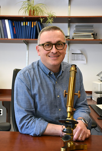
On a wooden desk sits 14 historical microscopes assorted in neat rows, gleaming a metallic gold in the office lighting. Manufactured between the 1800s-1900s in England and France, the instruments make up the personal collection of Dr. Italo Tempera, Associate Professor of Wistar’s Gene Expression & Regulation Program and Associate Director for Cancer Research Career Enhancement at Wistar’s Ellen and Ronald Caplan Cancer Center.

Tempera was interested in microscopy from a young age. “I started with biology when I was 13. I had a microscope at home,” he recalls. “I always wanted to collect and then at the beginning of the pandemic, I got the first one.” The microscope he refers to was made in 1830 in London and is the same kind of model that Darwin had. He also points out another model he finds most intriguing. Manufactured in France, the instrument was sold in Philadelphia from a shop on Chestnut Street, according to an inscription on the scope’s wooden box.

“We learn through our eyes. Microscopy is important because microscopes can reveal a lot of things we may not see through our experiments.” Tempera comments. “When you have a very powerful microscope image, you can really show and deliver your messages.” he continues.

The valuable knowledge that microscopes have given the scientific field – including how our cells work and are organized – emerged from these tools. In fact, Tempera and his colleagues recently used one from his collection (circa. 1890s – early 1900s London) to look at cancer tissue. “It was pretty good actually.” Tempera reveals in happy surprise.

The Horner Microscope

Tempera’s collection are not the only historical microscopes housed at The Wistar Institute. Nestled behind a glass display in the first floor Atrium is a microscope from 1830 made for Philadelphia anatomist William E. Horner. Made by Austrian optical instrument maker Simon Plossl, this microscope is still functional today. In 2017, Wistar’s Imaging Facility Managing Director James Hayden used the Horner microscope to image tumor cells and reconstructed skin from Wistar labs. He imaged the same samples with a modern Nikon microscope in Wistar’s Imaging Facility and compared the results.

“One of the things that surprised me was just how good it was. The fact that you could still separate cells and you could still see nuclei means that people 150 years ago could see the same stuff we can see now.” Hayden shares. “Even though technology has marched ahead, the basics haven’t changed much.”

What has changed with advances in microscopy is how images are used in scientific research. “The images are not just documentary anymore. They can be quantified. They become the data.” Hayden emphasizes.

Modern Microscopy at Wistar

Now many types of advanced microscopes exist, enhanced by the innovative technology of today’s world. From confocal microscopy to electron microscopy, the ability to magnify and quantify what would normally go unseen has expanded. Wistar research is helped tremendously by such powerful microscope technology at the Imaging Facility. For example, the Facility has imaged drug treatments on melanoma cells, stitched tissue images together, and recorded live cell behavior over mere milliseconds.

As a Shared Resource, Wistar’s Imaging Facility impacts diverse realms of research both internal and external to the Institute. “We have biological questions that need to be answered, so we take our existing technology, and we work with our industry partners to come up with new ways of imaging something to bypass the problem.” Hayden describes.

Whether it is virology or anatomy, biochemistry or chemical engineering, microscopy can be applied. Hayden says that it is the “investment in a facility that can keep up with the technology and more importantly, the hiring of people that can continually train others to support research” that makes microscopy at The Wistar Institute special.

Microscopes at Wistar

Highlighting Vaccine Research at The Wistar Institute Through the Penn-CHOP-Wistar Vaccine Symposium

From HIV to COVID-19, Wistar scientists are at the forefront of vaccine development. Read our recap of the recent Vaccine Symposium and the impactful research in progress at the Institute.

This past Monday, The Wistar Institute, University of Pennsylvania Perelman School of Medicine, and Children’s Hospital of Philadelphia held the Penn-CHOP-Wistar Vaccine Symposium. Hosted both in-person at the Smilow Center for Translational Research and online, the all-day event covered the history of vaccines and current vaccine research from the three sponsoring institutions.

Keynote speaker and Wistar professor emeritus Stanley Plotkin, M.D., is a prominent researcher who is known for the development of the rubella vaccine while he was a virologist at The Wistar Institute. Furthermore, his years of work helping in vaccine efforts for rabies, rotavirus, and cytomegalovirus have stimulated much innovation in the biomedical research community.
After giving a brief history of vaccines, Plotkin proclaimed “Vaccinology has taken off. … We are now in a golden age of vaccinology.”

The Symposium’s research presentations opened with Wistar’s Daniel Kulp, Ph.D., Associate Professor in the Vaccine & Immunotherapy Center, and his work on a novel COVID-19 nanoparticle vaccine. Amelia Escolano, Ph.D., Assistant Professor in the Vaccine & Immunotherapy Center, also spoke about her efforts investigating immunization strategies for HIV. Wistar’s Vaccine & Immunotherapy Center Director David B. Weiner, Ph.D., gave a summary of his research into the genetic delivery of vaccines, calling the innovation of vaccinology in Pennsylvania among these institutions “extraordinary”.

The current global pandemic has reinforced the need for scientific solutions and a deeper understanding of human diseases. It is the studies and ideas from research centers like The Wistar Institute and its colleagues that propel forward biomedicine. As keynote speaker Plotkin stated, “Pandemics have occurred throughout the history of humankind and will continue to do so in the future. Infectious diseases of humans will continue to happen. Therefore, we must act against them.”

The Wistar Institute Cancer Center to be Renamed with $10 Million Gift from Caplan Family to Advance Cancer Research

PHILADELPHIA — (March 8, 2022) — The Wistar Institute announces that Ronald Caplan, founder and president of PMC Property Group, Inc., and his wife Ellen have donated $10 million to Wistar’s prestigious National Cancer Institute (NCI)-designated Cancer Center, which will be renamed the Ellen and Ronald Caplan Cancer Center of The Wistar Institute.

The gift to Wistar, the nation’s first independent biomedical research organization, will foster Wistar’s world-leading scientists in their high-impact discoveries toward promising cancer therapies. The investment will also further Philadelphia’s growing recognition as a life science innovation hub.

“Despite recent progress, cancer is still a feared disease carrying enormous morbidity and mortality for patients and their families. Ellen and Ron’s vision is to change that: their generous gift will dramatically accelerate the pace of innovation and discovery at our renowned cancer research center,” said Dario C. Altieri, M.D., president and CEO of The Wistar Institute, director of the Ellen and Ronald Caplan Cancer Center, and the Robert and Penny Fox Distinguished Professor. “We are enormously grateful to the Caplans for their unwavering support and transformative leadership as we prevail in our mission to discover new ways to detect, treat, and defeat cancer.”

The Caplans have deep ties to Wistar. Ronald has been a member of the Institute’s Board of Trustees since 2009; in 2014 he and Ellen donated the 200-seat high-tech Sarah and Matthew Caplan Auditorium in the Robert and Penny Fox Tower – named for their children in the hope that cancer’s cure would be found in their lifetime.

Wistar’s Cancer Center became the nation’s first NCI-designated cancer center for basic research in 1972 and is among only seven such centers in the U.S. to continuously hold this distinction for a half century. The Cancer Center has a history of impactful advances in cancer genetics, cancer biology, tumor immunology, and virology, including the identification of genes associated with different tumor types, the development of monoclonal antibodies used to study pathways and proteins involved in tumor development, and contributions to improved cancer treatments and diagnostic tests.

Achieving NCI’s highest ranking of “exceptional” in the last two consecutive Cancer Center Support Grant renewal applications, the Cancer Center now becomes one of only a few named biomedical research cancer centers in the country.

The newly envisioned Ellen and Ronald Caplan Cancer Center will help transform the prevention and treatment of cancer by advancing fundamental and translational research into next-generation therapeutics. This will involve expansive recruitment of new leaders in cancer research focusing on key areas of treatment resistance, metabolic and cellular reprogramming, cancer systems biology and personalized anticancer strategies.

“Ellen and I are honored to invest in the source of scientific discovery – the scientists whose life-changing breakthroughs will continue to bring us one step closer to a future without cancer,” said Ronald Caplan.

“Wistar scientists are some of the most dedicated, brilliant people we have ever met. Thanks to their incredible work, Wistar is the one place where every time Ron and I leave, we feel inspired,” said Ellen Caplan.

Learn more about the Ellen and Ronald Caplan Cancer Center below.

Note: Photos are available on request.

###

The Wistar Institute is an international leader in biomedical research with special expertise in cancer research and vaccine development. Founded in 1892 as the first independent nonprofit biomedical research institute in the United States, Wistar has held the prestigious Cancer Center designation from the National Cancer Institute since 1972. The Institute works actively to ensure that research advances move from the laboratory to the clinic as quickly as possible. wistar.org.

Wistar’s Education & Training Team Visits Vaux High School to Connect on Life Science Careers

Wistar’s Drs. Kristy Shuda McGuire, dean of biomedical studies, and David Zuzga, associate dean of biomedical studies, spent an evening with students at Vaux Big Picture High School in north central Philadelphia to discuss all the STEM job opportunities available and for the taking here in the region.

Dr. Shuda McGuire spoke to Wistar’s education programs—available to high school students and upwards to postdoctoral fellows. She discussed how collaboration with academia and industry have made Wistar’s Biomedical Technician Training Program and Biomedical Research Technician Apprenticeship effective and successful vehicles to give students the needed skillset to enter coveted biomedical and biotechnical jobs. Then she imparted a little history on where and how Wistar fits into the dynamic regional life science hub we know today—from its beginnings as the first biomedical research institute in the nation, to how it has become a connector and engine for education in the burgeoning Philly biotechnology space.

Dr. Zuzga laid the groundwork on all the amazing science taking place in Philadelphia—from CAR T cell-trained assassins that became the first FDA-approved cell therapy to cure cancer in people who had no other options, to a new gene therapy company that created the first medicine to cure blindness. Dr. Zuzga drove home the point that the industry is booming in Philadelphia and students should set out and stake their claim in it. He emphasized that 10-years ago there were one or two cell and gene therapy companies and now there are more than 40 and the number is growing.

Basir Fulmore, a graduate of Cheyney University who is working in science, wrapped up the event and spoke to the students about his career trajectory. He talked about where he came from, the life lessons he learned in the process of attaining his science education and the true affinity he holds for science. He is a math and science teacher now but shared many stories of working in a Cheyney lab carrying out aquaponics—using fish to grow plants hydroponically. He compelled the students to enter science as more diversity is needed.

The Lower North Philadelphia CDC is a key partner, providing event coordination and sponsorship, as well as being an organization connector. Their friends at Give and Go Athletics brought a great group of students, who took part in the event and should be commended for taking two hours on a 70° day—after school—to learn more about science! It all seemed well worth it when they got to conduct their own scientific experiments and precipitated DNA from a solution. Eyes widened and smiled through masks. In those few moments you could see in each face that the magic of science homed in and made its first introduction.

VAUX Big Picture High School - Wistar Education Event

Women & Science: Pain, Itch, and Inflammation

Dr. Diana Bautista has dedicated her professional life to the science of literal human connection: our sense of touch.

A Howard Hughes Medical Investigator and professor of cell and developmental biology at University of California, Berkeley, Bautista runs a lab that conducts basic scientific research on touch, particularly itch, pain, and inflammatory diseases.

“Among the five senses, we really know the least about our sense of touch,” Bautista said. “This is surprising given how important touch is in our everyday lives.”

She points to pain as an example of why we need touch to stay healthy and safe. Acute pain, the feeling you get when you touch a hot frying pan, acts as a warning—it triggers a reflex to pull your hand back and prevent tissue damage. Likewise, inflammatory pain from a sunburn will prompt protective behaviors, like covering up before you go back outside, and learning behaviors, like proactively wearing sunscreen.

Chronic pain, however, serves no purpose. It’s like a siren that keeps ringing long after the fire has been put out. It’s a serious problem, afflicting more than 10 million Americans every year. “Which is why we’re working to understand the mechanisms underpinning touch and pain,” said Bautista. She and her colleagues are trying to determine why some people’s pain goes away when they recover from injury or disease, while other people suffer from chronic pain for the rest of their lives.

Bautista’s lab focuses primarily on the first step of feeling physical touch, which is called transduction. In this step, sensory neurons convert a physical stimulus, like the pressure of a handshake or the prick of a pin, into the electrical signals the nervous system uses to communicate. By identifying how these signals are triggered, Bautista hopes to identify new therapeutic targets for mediating pain.

“Our challenge is to discover new genes that underlie the persistence of chronic pain so we can develop new therapies that target pain at its source.” she said.

One gene her lab is researching is TRPM8, which is responsible for sensing cold and menthol. To verify this gene’s function, the scientists set up a mouse experiment. They created a small two-room chamber with an open doorway between the rooms. One room had a comfortably heated floor plate, and the other room had a very cold floorplate. When a regular mouse was put in the chamber, it avoided the cold side. However, when a mouse lacking the TRPM8 gene was placed inside, it traveled between the two chambers indiscriminately, demonstrating no sensitivity to the cold.

The discovery of this gene’s role in cold perception could help lead to treatment for patients suffering from cold allodynia (oversensitivity to cold), chemotherapy-induced neuropathy, and even certain types of migraines.

Another gene Bautista’s lab is studying is S1PR3. Rather than cold, this gene regulates sensitivity to mechanical pain. To test this, the researchers poked mice’s paws with a pin. Normal mice responded to this sensation 100% of the time, whereas mice lacking S1PR3 responded only 40% of the time. This finding is important because the gene is also expressed in human skin, as well as in a variety of other species.

Finally, Bautista’s lab is studying chronic itch, a condition that will affect one in ten people at some point in their lifetime. Like pain, acute itch is protective; it helps to ward off burrowing insects that might carry disease. However, chronic itch can be debilitating.

Historically, chronic itch has been associated with diseases of the integumentary (skin) and immune systems. However, Bautista’s lab has found that the nervous system interacts with the immune system in ways that can trigger inflammation from the skin all the way to the spinal cord. These interactions, which occur early in the sensation of itch, play a significant role in later chronic states of itch and can even go so far as to affect the onset of asthma.

“We’re really excited about this research,” said Bautista. “Very basic science can lead to the discovery of novel therapeutics.”