Author: The Wistar Institute

The Wistar Institute Announces the Appointment of Aleister Saunders, Ph.D., and Patrick Oates, Ph.D., to its Board of Trustees

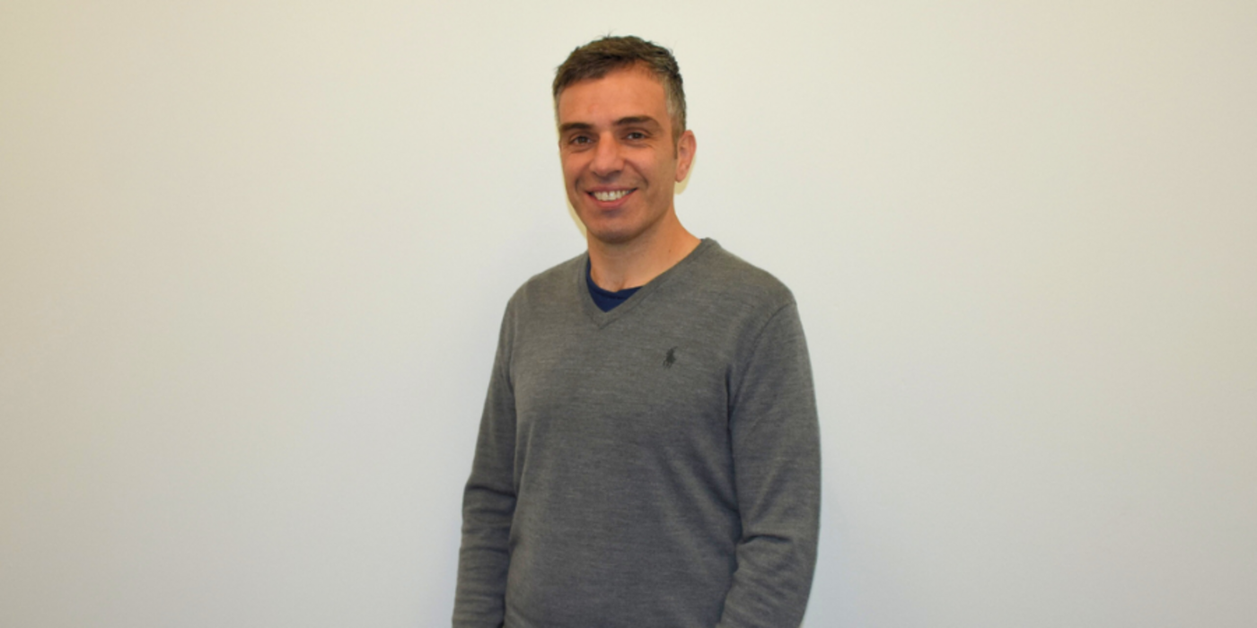
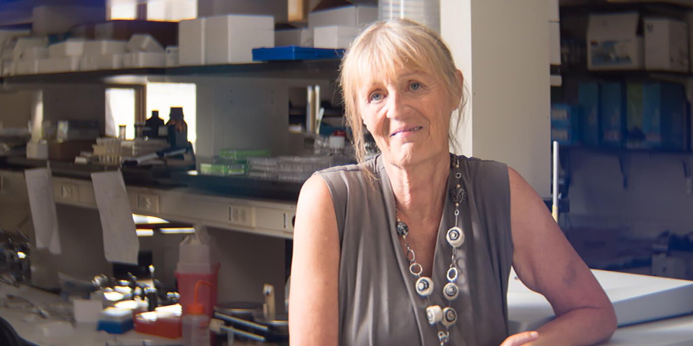
PHILADELPHIA—(Dec. 18, 2023)—The Wistar Institute, a global leader in biomedical research in cancer, immunology and infectious disease, is pleased to welcome Aleister Saunders, Ph.D., and Patrick Oates, Ph.D., to its Board of Trustees. The two new trustees bring a wealth of experience in biomedical research, technology transfer, and drug development to Wistar’s mission.

As Executive Vice Provost of the Office of Research & Innovation at Drexel University, Dr. Aleister Saunders is responsible for the strategic, compliance and grants management aspects of research as well as the licensing and commercialization of Drexel-created innovations. Prior to his current role, Saunders served as Drexel’s Senior Vice Provost for Research; Associate Dean for Natural Sciences Research and Graduate Education in the College of Arts and Sciences; Associate Department Head of the Department of Biology; and Director of the University’s RNAi Resource Center.

Dr. Saunders obtained his BS in biochemistry from the Pennsylvania State University and Ph.D. in biochemistry from the University of North Carolina at Chapel Hill. He also completed postdoctoral research fellowships at Harvard Medical School in functional genomics and later in genetics.

“As someone who has had a working relationship with The Wistar Institute for nearly 20 years, I’ve long been impressed with their focus and commitment to advancing meaningful discoveries,” explained Dr. Saunders. “From its history of therapeutic innovations to its workforce development and training efforts, I’m excited to see them continue leveraging new technologies and approaches to further contribute to Philadelphia’s thriving life sciences marketplace.”

Dr. Patrick Oates is the Senior Vice President of Business Development & Strategic Planning for EMSCO Scientific, Inc., where he manages partnerships with pharmaceutical companies and other life science entities to foster commercial growth. Prior to his role at EMSCO, Dr. Oates had numerous roles of increasing responsibility with a number of pharmaceutical companies, including GlaxoSmithKline, Aventis, Wyeth, and Cherokee Pharmaceuticals.

Dr. Oates completed his undergraduate training at Tuskegee University in Animal Science and received his doctorate degree in Physiology & Molecular Biology at Howard University in Washington, DC.

“The Wistar Institute has long been a leader in Philadelphia’s biomedical research landscape, and their commitment to educating the next generation of biomedical professionals is advancing equitable career opportunities in this growing market,” said Dr. Oates. “Their work is vital to continue diversifying the STEM workforce and I look forward to joining with their leadership team to continue those efforts.”

“We’re extremely fortunate to add the expertise of two inspirational and accomplished leaders like Dr. Saunders and Dr. Oates to our leadership team” said Dario Altieri, M.D., president and CEO of The Wistar Institute. “They bring with them not only a deep understanding of The Wistar Institute and its vision, but also a commitment to excellence and a passion for innovation that dovetail perfectly with Wistar’s mission. We look forward to working with them to further advance Wistar Science and continue our record of bringing forth transformative biomedical discoveries that improve the future of human health.”

Learning to Listen to Our Cells: 20 Years of The Society for Melanoma Research with Dr. Meenhard Herlyn

Meenhard Herlyn, D.V.M., D.Sc., founded the Society for Melanoma Research 20 years ago at Wistar. The Society’s annual Congress just celebrated its 20th anniversary and presented Dr. Herlyn with the Founders Award, an honor commemorating his work in creating an organization that has been integral to the advancement of melanoma research.

We were told that, before you received the Founders Award, it was supposed to be a surprise. Did they succeed in surprising you?

Oh, they absolutely succeeded. I had not been expecting anything at all. I am not really a surprise person, but I was quite moved by this gesture. Everyone got up — it was a real honor. I felt so energized by the moment that I gave a little spontaneous pep talk.

A pep talk? What did you say?

I spoke about the SMR mission, which is bringing together clinicians and researchers against melanoma. The idea is to bring together the strengths of research and clinical practice — but this can only happen when we are transparent and open. Throughout my career, I’ve found that progress is made by sharing: sharing data, sharing methods, sharing tools — everything! I know that, sometimes, scientists might not show all their data publicly, out of administrative concerns or something — but to me, that is a big no-no, especially in an organization like the SMR. We are proud to be transparent because that is how we move forward.

Do you think that the openness in your field has increased over the years?

Oh, of course; the openness has expanded tremendously, and I’m proud to say that our field stands out for its collaborative approach. We now almost have the opposite problem, where so much data accrue thanks to what we call “omics” technologies (as in, genomics, proteomics, etc.). These datasets require specialized bioinformatics knowledge to interpret because of their complexity and sheer size. My lab has approximately 80 terabytes of data, and I estimate that by this time next year, that quantity will have doubled or even tripled. We have great resources, yes. But we are also searching for needles in haystacks.

Our cells speak to us. They tell us at each and every step how they feel, what they do, and with whom they interact. As researchers, it is our job to listen to what our cells are saying, but that listening is quite difficult. We do our best with modern technology to gather all these signals from RNA, DNA, proteins — and all of this for each cell, tens of thousands of them. It’s like a big crowd, roaring. To listen to the cells, we must sort out the noise from the useful information to see which genes and proteins are talking, telling us what’s wrong.

What inspired you to start the SMR?

20 years ago, we had two communities: we had the researchers, busy in the lab, looking at data; and we had the clinicians, diagnosing and treating patients while relying primarily on clinical experience. At the time, I was working with a very gifted pathologist, Dr. Wallace Clark, and we were investigating melanoma, a field where there was very little progress being made; the treatments worked maybe 2% of the time. While working on the project and trying to obtain samples, I realized how big the gap between researchers and practitioners was. The two groups were each cooking their own soup, not talking to each other. So I figured that bridging the gap was essential for making any progress at all, because having the clinic separate from the lab just wasn’t working. That’s where the idea for the SMR came from.

It wasn’t SMR at first, though. We were the Noreen O’Neil Foundation for Melanoma Research. Noreen was a melanoma patient who saw that there was very little melanoma research, and she also recognized that this had to change. She spoke with her clinician, Dr. DuPond Guerry, and we began the work of putting together the organization. Unfortunately, Noreen died of her illness, and so her sister Kate O’Neil took over. With their generosity, we had $50,000 in the bank, which gave us two options. The first was awarding grants, of which we could award two and then the money would be gone. Or, we could use the money to set up a meeting for melanoma researchers and clinicians. So we got to work finding a space, inviting speakers, making sure that the lab perspectives were balanced with the clinical perspectives — and the Congress was born.

The first SMR Congress was an enormous success. We decided to hold it in a new city every year after, to keep it fresh and exciting, but having the 20th anniversary back in Philadelphia where it started was a nice touch.

How far have you come, and how much further do you have to go?

We have many miles to go yet, but I’m proud to look back on the progress we’ve made. Not only scientifically, but also in terms of widening the community. Researchers and clinicians work together against melanoma now, sharing their insights with each other — that shows the power of openness in science. I’m especially proud of the work that my colleague Dr. Jessie Villanueva is doing for Diversity, Equity, and Inclusion in science. Science only works when people come together to solve problems; there can be no room for exclusion or bias because everyone’s insight is needed to advance the field.

On the other hand, we need to keep pushing forward. With the frequency of melanoma cases rising, research is more important than ever; keep in mind that, for all the advances the field has seen, we can still only successfully treat 50% of patients long-term. When every other patient eventually dies of their illness, that tells you just how vital it is to keep up the work. Only by bringing more talent into the fold across disciplines will we be able to sustain progress against this cancer.

The Wistar Institute Recruits Dr. Filippo Veglia to the Ellen and Ronald Caplan Cancer Center

PHILADELPHIA—(Dec. 13, 2023)— The Wistar Institute, an international biomedical research leader in cancer, infectious disease, immunology, and vaccine development, is pleased to announce the recruitment of Filippo Veglia, Ph.D., to the Ellen and Ronald Caplan Cancer Center, where he joins Wistar’s Immunology, Microenvironment and Metastasis Program as an Assistant Professor. 

As a researcher who studies glioblastoma, the most lethal form of brain cancer, Dr. Veglia investigates how glioblastoma functions at the foundational level in hopes that he can uncover potential weaknesses that, once identified, could be targeted. Glioblastoma’s lethality comes from its aggression and sheer difficulty to treat; in an organ as fragile as the brain, cancer is even more dangerous, and glioblastoma spreads quickly. But Dr. Veglia’s expertise in myeloid-derived immune cells — cells from bone marrow that perform immune functions — has allowed him to probe the tumor microenvironment of glioblastoma to search for new potential treatment targets. By analyzing how improperly functioning myeloid-derived immune cells enable glioblastoma tumors, Dr. Veglia hopes to find ways of stopping their cancerous influence. His research is at the intersection of cancer metabolism, cancer immunology, and the tumor microenvironment of glioblastoma. 

“We’re happy to welcome Filippo to Wistar’s faculty, where he will contribute his expertise in tumor immunology to a thriving culture of immunology research,” says Dario Altieri, M.D., Wistar president and CEO, director of the Ellen and Ronald Caplan Cancer Center, and the Robert and Penny Fox Distinguished Professor. 

“Many scientists at Wistar investigate the tumor microenvironment and immune cell dysregulation as part of their research, and Filippo stands to contribute his perspectives from his work in glioblastoma. In fact, I should say that we’re welcoming Filippo back to Wistar, because he trained here as a staff scientist years ago. We were glad to have him then, and he has become a promising young investigator who is pursuing exciting work that combines epigenetic, metabolic, and immunological approaches to cancer. We’re even more glad to have him now as a faculty member.” 

Wistar Scientists Enhance Cell-Based Therapy to Destroy Solid Tumors

PHILADELPHIA—(Dec. 13, 2023)—Wistar researchers successfully tested a simple intervention that could unlock greater anti-tumor power in therapies that use T cells — an approach known as “cell-based therapy,” which uses specially designed T cells to fight cancer. Led by Dr. Hildegund C.J. Ertl — a professor in The Wistar Institute’s Vaccine & Immunotherapy Center — the team has proven an exciting concept: that the common cholesterol drug fenofibrate can boost T cells’ ability to destroy human tumors, as described in their new paper, “Treatment with the PPARα agonist fenofibrate improves the efficacy of CD8+ T cell therapy for melanoma,” published in Molecular Therapy Oncolytics.

CD8+ T cells work very well in fighting liquid tumors, but for solid tumors like melanoma, the cell-based therapy approach can stall due to the physical structure of the cancer. The T cells infiltrate the tumor, but the cancer adapts and saps the T cells’ energy by hijacking the form of metabolism that the T cells use: glycolysis, which turns sugar into energy. Without energy, the T cells first lose functions and then die, and the cancer continues to grow.

But Dr. Ertl’s team has been able to circumvent this problem by forcing T cells to use a different energy source than glucose. They used fenofibrate because, as a cholesterol-lowering compound, the drug is a PPARα agonist. When PPARα is upregulated, cellular metabolism is switched from glycolysis to fatty acid oxidation, or FAO. This mechanism works to improve cholesterol levels in human patients, but for Dr. Ertl’s purposes, the fenofibrate-induced switch to FAO provided T cells with a form of energy that cancer couldn’t exploit — which is how Dr. Ertl proved that fenofibrate has been able to boost the killing power of T cells deployed against cancerous cell lines.

In this paper, the authors wanted to see whether this kind of cancer-killing improvement would have similar effects when deployed against not just cancer cell lines but solid human tumor fragments — a more challenging proposition. The group treated T cells with fenofibrate, and the hypothesis held: Dr. Ertl’s team watched the T cells treated with fenofibrate survive longer and kill more cancer in preclinical models with human solid tumor masses than the T cells that didn’t receive the treatment.

“Treating T cells with fenofibrate before using them as a cancer treatment flips a switch of sorts in their metabolism,” said Dr. Hildegund Ertl. “Once that switch is flipped, T cells can destroy the cancer much more effectively. And we’ve confirmed that this holds for larger human tumor masses.”

As a result of these findings, Dr. Ertl and her team think this intervention shows great promise for future anti-tumor therapies. “Melanoma is the most dangerous form of skin cancer. Anything we can do to chip away at the cancer and destroy more of it — even a simple pre-treatment step like this one — can make a world of difference.”

Co-authors: Mohadeseh Hasanpourghadi, Arezki Chekaoui, Sophia Kurian, Robert Amrose, Wynetta Giles-Davis, Amara Saha, and Hildegund C.J. Ertl of The Wistar Institute; Raj Kurupati of The Wistar Institute and The Janssen Pharmaceutical Companies of Johnson & Johnson; and Xu Xiaowei of the Hospital of the University of Pennsylvania

Work supported by: Department of Defense grant number W81XWH-19-1-0485 CA180191.

Publication information:Treatment with the PPARα agonist fenofibrate improves the efficacy of CD8+ T cell therapy for melanoma,” published in Molecular Therapy Oncolytics.

Filippo Veglia, Ph.D.

Assistant Professor, Immunology, Microenvironment and Metastasis Program, Ellen and Ronald Caplan Cancer Center

Veglia’s research focus is the study of molecular and metabolic perturbations that drive the reprogramming of myeloid cells in the brain tumor microenvironment, with a final goal to identify novel therapeutic strategies to target immunosuppressive myeloid cells.

Filippo Veglia received his Ph.D. in Immunology and Applied Biotechnology from Tor Vergata University in Rome (Italy) and did his postdoctoral work at Wistar in the laboratory of Dmitry Gabrilovich at Wistar. In 2020, he joined Moffitt Cancer Center as an Assistant Member in the Department of Immuno-Oncology and as a Member of the Neuro-Oncology Program. In December 2023, Filippo joined Wistar as an Assistant Professor in the Immunology, Microenvironment and Metastasis Program. He is an expert in myeloid cells, tumor immunology, and metabolism, and he has a proven track record of publications studying the metabolic regulation of myeloid cell functions in cancer as well as exploring novel strategies for their therapeutic targeting (Nature Communications, Nature, and Journal of Experimental Medicine).

View Publications

The Veglia Laboratory

215-898-3926

fveglia@wistar.org

The Veglia Laboratory

Glioblastoma (GBM) is the most aggressive and lethal form of brain cancer, which is characterized by a profound immunosuppressive tumor microenvironment (TME) that prevents the induction of effective anti-tumor immunity, thereby rendering these tumors highly resistant to immunotherapy.

Myeloid cells, which are the largest immune cell components of GBM tumors, are highly plastic and heterogenous in the TME. In response to the local clues provided by the TME, myeloid cells undergo an extensive remodeling of core energy metabolism and acquire

an entire spectrum of metabolic and functional profiles. Such extensive metabolic adaptation is critical for myeloid cell functions in the TME. Our lab focuses on the study of the TME-driven mechanisms responsible for the reprogramming of myeloid cells towards cells with immunosuppressive and pro-tumoral functions. Our current effort is to identify metabolic and molecular pathways shared by different immunosuppressive myeloid cells to enable the simultaneous targeting of these cells in the brain tumor TME, with final goals to restore anti-tumor immunity and boost the efficacy of immunotherapy.

Staff
  • Staff Scientist

    Alessandra De Leo, Ph.D.

  • Postdoctoral Fellows

    Barbara Peixoto, Ph.D.
    Alessio Ugolini, Ph.D.

Available Positions

Postdoctoral fellow and research assistant positions are available in the Veglia Laboratory. Candidates should have recently received or be close to obtaining their Ph.D. degree or equivalent (for postdoc) or B.S. degree or equivalent (for RA) and have a strong background in in immunology, tumor microenvironment, and molecular biology. Interested applicants are invited to email: fveglia@wistar.org

Research

Project #1: Metabolic-driven post-translational modifications supporting the suppressive and pro-tumoral function of tumor-associated macrophages and neutrophils in brain tumors

Project #2: Brain tumor-driven mechanisms of phenotypic and functional heterogeneity of neutrophils

Project #3: Role of microglia during brain tumor progression

Project #4: Metabolic mechanisms of trained immunity in brain tumors

Staff
  • Staff Scientist

    Alessandra De Leo, Ph.D.

  • Postdoctoral Fellows

    Barbara Peixoto, Ph.D.
    Alessio Ugolini, Ph.D.

Available Positions

Postdoctoral fellow and research assistant positions are available in the Veglia Laboratory. Candidates should have recently received or be close to obtaining their Ph.D. degree or equivalent (for postdoc) or B.S. degree or equivalent (for RA) and have a strong background in in immunology, tumor microenvironment, and molecular biology. Interested applicants are invited to email: fveglia@wistar.org

Lab in the News

Selected Publications

  • Analysis of classical neutrophils and polymorphonuclear myeloid-derived suppressor cells in cancer patients and tumor-bearing mice

    Citation: Veglia F, Hashimoto A, Dweep H, Sanseviero E, De Leo A, Tcyganov E, Kossenkov A, Mulligan C, Nam B, Masters G, Patel J, Bhargava V, Wilkinson P, Smirnov D, Sepulveda MA, Singhal S, Eruslanov EB, Cristescu R, Loboda A, Nefedova Y, Gabrilovich DI. Analysis of classical neutrophils and polymorphonuclear myeloid-derived suppressor cells in cancer patients and tumor-bearing mice. J Exp Med. 2021 Apr 5;218(4):e20201803. doi: 10.1084/jem.20201803. PMID: 33566112; PMCIDPMC7879582.

  • Myeloid-derived suppressor cells in the era of increasing myeloid cell diversity

    Citation: Veglia F, Sanseviero E, Gabrilovich DI. Myeloid-derived suppressor cells in the era of increasing myeloid cell diversity. Nat Rev Immunol. 2021 Aug;21(8):485-498. doi: 10.1038/s41577-020-00490-y. Epub 2021 Feb 1. PMID: 33526920; PMCIDPMC7849958.

  • Polymorphonuclear myeloid-derived suppressor cells limit antigen cross-presentation by dendritic cells in cancer

    Citation: Ugolini A, Tyurin VA, Tyurina YY, Tcyganov EN, Donthireddy L, Kagan VE, Gabrilovich DI, Veglia F. Polymorphonuclear myeloid-derived suppressor cells limit antigen cross-presentation by dendritic cells in cancer. JCI Insight. 2020 Aug 6;5(15):e138581. doi: 10.1172/jci.insight.138581. PMID: 32584791; PMCIDPMC7455061.

  • Fatty acid transport protein 2 reprograms neutrophils in cancer

    Citation: Veglia F, Tyurin VA, Blasi M, De Leo A, Kossenkov AV, Donthireddy L, To TKJ, Schug Z, Basu S, Wang F, Ricciotti E, DiRusso C, Murphy ME, Vonderheide RH, Lieberman PM, Mulligan C, Nam B, Hockstein N, Masters G, Guarino M, Lin C, Nefedova Y, Black P, Kagan VE, Gabrilovich DI. Fatty acid transport protein 2 reprograms neutrophils in cancer. Nature. 2019 May;569(7754):73-78. doi: 10.1038/s41586-019-1118-2. Epub 2019 Apr 17. PMID: 30996346; PMCIDPMC6557120.

  • Lipid bodies containing oxidatively truncated lipids block antigen cross-presentation by dendritic cells in cancer

    Citation: Veglia F, Tyurin VA, Mohammadyani D, Blasi M, Duperret EK, Donthireddy L, Hashimoto A, Kapralov A, Amoscato A, Angelini R, Patel S, Alicea-Torres K, Weiner D, Murphy ME, Klein-Seetharaman J, Celis E, Kagan VE, Gabrilovich DI. Lipid bodies containing oxidatively truncated lipids block antigen cross-presentation by dendritic cells in cancer. Nat Commun. 2017 Dec 14;8(1):2122. doi: 10.1038/s41467-017-02186-9. PMID: 29242535; PMCIDPMC5730553.

View Additional Publications

Continue reading

Wistar’s Inaugural Diversity, Equity & Inclusion (DEI) Survey Helps Chart a Path Forward 

Diversity, Equity and Inclusion has historically been a priority for The Wistar Institute. Creating a diverse and inclusive environment not only attracts top-tier talent, but it also supports innovation, creativity, and a commitment to collaboration amongst existing staff.

Since it was established in 2021, The Wistar Institute’s Inclusion, Diversity, and Equity (W-IDE) council, under the leadership of Dr. Jessie Villanueva, has been focused on further enabling an inclusive culture. As part of the effort, the Wistar Institute, recently issued a DEI survey to gather its staff’s perspectives on enabling a culture of equity, diversity and inclusion as well as establish a baseline for further evaluation long-term. Feedback from the W-IDE council was integral in the anonymous survey that was developed and administered by an independent consultant, ADI.

With a response rate of approximately 71% — a rate that surpassed many peer organizations at this phase — Wistar staff sent a clear signal that they value DEI initiatives and are engaged in the efforts. And the overarching results were encouraging: a strong majority (nearly 97% of respondents) agree that Wistar fosters a supportive, welcoming, and inclusive environment.

The survey included a total of 39 questions organized across six different categories. Responses were rated on a scale of one to eight, with a rating of “1” being the lowest and “8” being the highest.

“We want to be in the range of six to eight on the scale,” explained Dr. Villanueva during a presentation of the results. “Every single category that was surveyed had a score higher than six. That’s something that we can all be proud of.”

The category that had the highest score was manager and PI feedback. “That indicates that leaders in the Institute are being very effective, fostering an inclusive and diverse environment,” said Dr. Villanueva.

Another area of strength for Wistar is providing support opportunities for those who need it: The survey revealed that 90% of staff believe that they have at least one colleague that they can reach out to for support.

Vice president of Human Resources Jo-Ann Mendel emphasized that DEI efforts require broad support and collaboration to achieve the best outcomes. “This is not something that we can do ourselves. We need to build partnerships and make sure we all contribute to the success of these plans,” she explained. “Together we will be able to accomplish our goals.”

Moving forward, the W-IDE council is partnering with departments and stakeholders throughout Wistar to address some areas for improvement revealed by the survey. For instance, the results suggested that people felt that Wistar does not properly recognize and give credit to those who contribute to the success of the institution. “We heard you and we plan to develop better strategies to recognize those folks,” said Dr. Villanueva.

“This survey is just the beginning,” commented Dr. Villanueva. “We have established additional channels to provide for all members of the institute to give us additional feedback.”

For more information, or if you have any questions or comments about the survey or our plans to enhance diversity, please feel free to contact the W-IDE council at WI-IDE@wistar.org or leave us a comment online.

For Wistar researcher Dr. Gundi Ertl, time away from the lab is best spent with furry — and feathered — friends.  

Managing a lab involves a complex orchestration of tasks: running experiments, writing grants, engaging staff, managing the research budget, and answering emails. With many tasks to juggle, you wouldn’t be wrong to think that when the day ends or the weekend rolls around, perhaps a Principal Investigator (PI) would want a break or some time alone.

For Dr. Gundi Ertl, professor in Wistar’s Vaccine & Immunotherapy Center, an entirely different sort of team is waiting for her when she arrives home, but one that brings her peace: two hulking Great Danes, three cats, and one ill-behaved African Grey parrot, all clamoring for her attention, and maybe a treat.

“I’ve had pets all my life,” she explains, sharing the details of her childhood growing up in Germany with cats. “My family owned a company that made cloth. The factory was on a big property and there were many sheds. So we needed cats to manage small pests, and I was always allowed to have one.”

After finishing medical school in Germany, she got her own cat. But a year later, after landing a fellowship opportunity in Australia, she was forced to leave the cat with her mother. Not to be deterred, though, Dr. Ertl made her way to Australia, found temporary housing in a hostel and immediately got herself a Siamese cat. “That cat eventually came with me to Boston,” she explained.

That cat is the entire reason why Dr. Ertl became drawn to her beloved Great Danes. 

Following her fellowship in Australia and another at the University of Minnesota, Dr. Ertl was recruited by Harvard University, and she and her cat moved to Boston. While living there, she made an unplanned visit to the vet to tend to the ailing Siamese. It was in the waiting room of the vet clinic that Ertl first saw what would become her preferred dog. 

“The cat was seated on my lap, and she was clearly uncomfortable,” she recalls.  “In walks this man with a black Great Dane. I was mesmerized — it was the most beautiful animal I’ve ever seen.”

Since that day, Dr. Ertl has owned seven Great Danes, and currently has one 9-year-old male, Papageno, who is 180 lbs., and a 3-year-old female named Black Magic, who weighs in at 140 lbs. “There’s no pretension. They can be obnoxious, but by and large, the dogs are always happy to see me. They’re fun to watch, and if they complain you can ignore them,” she says laughing. 

Dogs, of course, have a mind of their own and ignoring them is not always an option. Dr. Ertl tells one such story from her time in Boston, when she decided to train her dog in an obedience program. 

“There is one exercise where you make your dog sit, you walk away 20 or 30 feet, and the dog must wait until you call it. We were in a large open area, next to some fenced in areas,” she describes. “I called my dog and she started running toward me at 100 miles an hour, and just before she reached me she veered off, jumped over the fence and into an area with a Golden Retriever.” After some choice words from the other dog’s owner, Gundi and her dog were excused from the program. 

On the feline side, Gundi now has three cats in her house: a Siamese named Wendolin, an Egyptian Mau named Mozart, and a 15-pound Maine Coon named Maximilian, all of whom coexist comfortably with the dogs. “The Maine Coon can be amazingly obnoxious, but she is very beautiful.”

Then there’s the African Grey Parrot named Matilda that Gundi adopted 12 years ago from a rescue, who has a bit more of a surly personality.

“When I picked her up from the rescue, she looked at me and said, ‘Oh no, I’m not going with you.’ And I decided, ‘You bet you are!’ It was sort of a challenge,” she says. “She’s not a very friendly bird. If you get too close to her cage she’ll bite, so everyone stays away.” 

“When I first got the bird, her name was Spanky,” she continues. “I decided I hated that name, so I called her Matilda. But now every evening I say ‘good night, Matilda’ and she responds with ‘Good night, Spanky bird.’ Twelve years later and we are still fighting that issue.”
But challenges are not something that Dr. Ertl shies away from. After nearly 36 years as a Wistar researcher — and as a female scientist at the forefront of vaccine research — she’s learned to stand her ground. “I have a reputation of saying what I think,” she explains. “As women, we can’t be intimated and must stop being shy and subservient.” 

During her career she’s also seen things improve for rising female scientists. “20 or 30 years ago I would be the only female speaker to get invited to conferences, and that happened over and over again,” she recalls. “That has stopped. Now more women are getting invited, so it has gotten better.” 

So what does the future hold for Dr. Ertl?

“I love science. I love my people,” she says of her lab team at Wistar. “I also co-founded a company that has future potential. And I have family in Morocco. So I’ve thought about moving to Morocco where I can continue consulting. I’m going to stay active, not sit on my hands.”

And if she leaves the country, would the pets go with her?

“Yes. Absolutely, though I’m not so sure about the parrot. Some countries have strict import rules about animals, so we’ll have to figure that out.”
But in the end, even an ill-mannered parrot is a beloved companion through and through. 

How the Wistar Flow Cytometry Facility Came to Be

Jeffrey Faust keeps a toolbox next to his office. It looks like something you might find in a garage: screwdrivers, Allen wrenches (standard & metric), wire caps, the indispensable duct tape, and so on. But Jeffrey doesn’t tinker with cars or ham radios — his tools are for adjusting and maintaining flow cytometry equipment with six-figure price tags. That doesn’t faze Mr. Faust, though; he’s been doing this for more than 40 years.

Jeffrey Faust, M.B.A., is the Managing Director of The Wistar Institute’s Flow Cytometry Core Facility, where the days are spent sorting, counting, and analyzing cells. Scientists from around the institute — and even beyond; as a Core Facility, the resources here are commercially available to external parties — depend on Jeffrey and his team to turn biology into data that can be interpreted.

Mr. Faust’s domain is strictly all-business: a set of fluorescently lit rooms that would be unassuming if not for the array of massive whirring machinery that gives the facility a space-age aura. Tools, parts, and sets of instructions line the walls, and the monitors attached to the active machines display live feeds of data as it is collected, displayed, and analyzed.

Having started the facility from scratch, Jeffrey has an impeccable command of all things flow cytometry. He first walked through Wistar’s front doors in 1977, fresh out of the University of Pennsylvania with a degree in natural sciences and hoping only for some form of gainful employment during the 70s’ economic malaise. Hired as secretary, the young Jeffrey soon became a lab technician working with mRNA — until his PI unexpectedly and tragically died, and Jeffrey was thrust into the role of lab manager.

In late 1980, Wistar, eager to stay up to date on the latest in scientific instrumentation, purchased its first flow cytometer; the nascent technology was still new to the world of biological research, and Wistar needed someone who could operate it. An offer was made, and Jeffrey Faust was soon enrolled in a three-week training course to understand how the machine worked — Wistar had found its Director of Flow Cytometry.

So what does a flow cytometer do? A flow cytometer aims a laser of a specific wavelength at a steady stream (or flow) of cells that have been stained with a special chemical called a fluorochrome — which, when excited by light of wavelength x, will emit light with wavelength y. When each cell flows through the laser’s path, the fluorochromes emit certain wavelengths of light, which, after going through a series of optical filters, then hit a detector that converts those emissions into electrical pulses. Those pulses are converted into digital signals that are sent to an attached workstation. This process allows flow cytometers to convert thousands and thousands of signals from the flow of raw cells into data that can be visualized and analyzed.

Because different fluorochromes can bind to different parts of a cell, they can be used to measure different properties. Let’s say you want to know how many cells in a given population are alive after a treatment. You can treat the cells with a fluorochrome that’s known to bind to dead cells but not living ones. Then you process the cell population with the flow cytometer, and for every fluorochrome emission signal you see on the screen, you know you have a dead cell. A similar process allows for the precise determination and quantification of antigen expression.

With the advancement of fluorochrome technology over the years, scientists have been able to use this technology to quantify a variety of cell characteristics in bulk populations; if a phenomenon can be tagged with a fluorochrome, then a flow cytometer can measure it. The Wistar Institute Flow Cytometry Facility now has instruments capable of detecting up to 48 total fluorochromes.

Back when Jeffrey Faust installed the very first of Wistar’s flow cytometers, the technology was still very much in its infancy — limited laser wavelengths and even fewer fluorochromes. And while today’s advanced FC equipment is designed with the user in mind, the first-gen flow cytometers were aggressive in their user-unfriendliness; the picture Jeffrey has of the original machine and its computer resembles nothing so much as a knob- and dial-studded sarcophagus and an antique arcade game, respectively.

“We were really just figuring this stuff out ourselves because we were right there at the beginning of this technology,” says Jeffrey, looking back on the history of the facility. “I’d call up someone halfway across the country who used the same machine, and we’d compare notes. Because we had to; otherwise, we weren’t going to make any progress.”

The machines got bigger, faster, and easier to use, but Jeffrey kept current. In his day, he’s worked with scores of scientists — even Nobel Prize winners, including Wistar’s own Dr. Peter Doherty (1996 Nobel Prize Winner, Physiology or Medicine). The discovery of a monoclonal antibody to detect natural killer cells (NK cells) was an early major discovery in the Wistar Flow Cytometry Facility. “There had not been monoclonal antibodies available that targeted NK cells. At the time, Wistar was working to develop and isolate the very first monoclonal antibodies, and we kept noticing this little shoulder in our flow cytometry data. As that lab, which was Trinchieri and Perussia, dug into it, they narrowed the data down — until we realized that they were able to quantify NK cells for the first time.”

In his time as facility director, Jeffrey has managed and hired 16 technicians to assist him in the facility — eight of whom moved on to manage their own facilities in academia and industry. Wistar’s facility continues to grow, with Jeffrey having helped incorporate spectral flow cytometry as well, which uses additional sensitive wavelength detectors to capture full-spectrum flow data across several parameters. To manage the ever-growing demands for flow cytometry data, Mr. Faust and his staff continue to train and assist users, who learn how to process millions and millions of cells a day with the precision necessary to keep the research moving forward accurately and efficiently.

How have the 40+ years affected Mr. Faust? Hardly at all, it seems. The “guts” of his first machine are still in the office, but the real energy of this facility is in its current operations, not its past. Flow-cytometry data, as it’s being collected and graphed, looks like a real-time pointillism piece, both patterned and strangely abstract. To the non-expert, it’s a mesmerizing sight; to Jeffrey Faust, it’s what a good day’s work looks like. “You see that?” he says, pointing to a line on the screen of an instrument in use. “That’s where the dead cells are excluded.” There are seemingly thousands of dots appearing by the second. And the machine — perfectly adjusted and expertly tuned — keeps counting.

Wistar Scientists Identify Esophageal Cancer Biomarkers

Dr. Noam Auslander and authors trained a neural network to identify cancer risk from microbes.

PHILADELPHIA—(Dec. 5, 2023)—Wistar scientists have developed a new tool that can help identify cancer-associated microbes by using machine learning technology. Under the leadership of Dr. Noam Auslander — assistant professor in the Ellen and Ronald Caplan Cancer Center’s Molecular & Cellular Oncogenesis Program — the group has analyzed short read RNA-sequencing data to detect biomarkers for esophageal carcinoma, or ESCA. Their paper, “Microbial gene expression analysis of healthy and cancerous esophagus uncovers bacterial biomarkers of clinical outcomes,” was published in International Society for Microbial Ecology Communications.

Tumor microenvironments are often analyzed using RNA sequencing, or RNAseq, which identifies mRNA in a population of cells to find which genes are being expressed. Theoretically, RNAseq data can reveal the expression of microbial genes in cancerous tissue, which could help to identify microbiome shifts that might be playing a role in the cancer’s development. But RNAseq “reads” — the physical lengths of genetic data that correspond with gene expression — are often quite short, posing a challenge for classifying them into diverse microbial genetic origins. Assembling the short RNAseq reads into longer contiguous segments that can be associated with a vast array of potential origins — be they human, viral, or bacterial — to identify specific microbes whose expression correlates with ESCA is computationally challenging.

That’s where Dr. Auslander and her group decided to intervene by training a convolutional neural network, a type of machine-learning technology that can be taught to train itself to accurately assess large quantities of data. The team, using large publicly available datasets of characterized short-read data, trained the network to sort short-read RNAseq data by its likely origin: human, viral, or bacterial. Their model sought to pare down the number of short reads that would need to be assembled for identification, which would reduce the computational load of screening for microbial influences in cancer tissue.

Once the model was trained, its sorting capabilities allowed the group to selectively analyze ESCA tissue for reads of microbial origin and compare those data with apparently healthy esophageal tissue. Auslander’s team found several instances of microbial expression present in ESCA with significantly less incidence in apparently healthy esophageal tissue.

In particular, they found that nearly half of the microbial genes over-expressed in cancer originated from bacteriophages, which are viruses that infect bacteria; this finding may indicate that viruses infecting microorganisms within the tumor microenvironment facilitate ongoing cancerous gene expression.

The team also identified patient outcome predictors amid the data. Bacterial iron-sulfur proteins were found to impact human genes involved in ferroptosis — a type of cell death pathway that’s modulated by iron — which predicted poor prognosis in ESCA patients. Microbial genes involved in mitochondrial reprogramming were also found to predict ESCA patient prognosis.

“By building on our previous work, our team has successfully leveraged machine learning to dive deeper into what’s going on inside cancer,” said Dr. Auslander. “While it’s always important to remember that correlation does not equal causation, the associations we’ve been able to find between certain microbial genes and ESCA will allow scientists to further understand the mechanics of esophageal cancer — which is the first step in stopping it.”

Co-authors: Daniel E. Schäffer of Carnegie Mellon University, The Wistar Institute, and the Massachusetts Institute of Technology; Wenrui Li of The University of Pennsylvania; Abdurrahman Elbasir and Dario C. Altieri of The Wistar Institute; Qi Long of The University of Pennsylvania; and Noam Auslander of The Wistar Institute.

Work supported by: National Cancer Institute grant numbers R00CA252025 and P30-CA016520 and National Institute on Aging grant number RF1-AG063481.

Publication information: “Microbial gene expression analysis of healthy and cancerous esophagus uncovers bacterial biomarkers of clinical outcomes,” published in International Society for Microbial Ecology Communications.

###

The Wistar Institute is the nation’s first independent nonprofit institution devoted exclusively to foundational biomedical research and training. Since 1972, the Institute has held National Cancer Institute (NCI)-designated Cancer Center status. Through a culture and commitment to biomedical collaboration and innovation, Wistar science leads to breakthrough early-stage discoveries and life science sector start-ups. Wistar scientists are dedicated to solving some of the world’s most challenging problems in the field of cancer and immunology, advancing human health through early-stage discovery and training the next generation of biomedical researchers. wistar.org

Wistar President Dario Altieri, M.D., recognized as one of Philadelphia’s Most Admired CEOs

PHILADELPHIA—(Dec. 4, 2023) — Dario C. Altieri, M.D.The Wistar Institute’s president and CEO, director of its Ellen and Ronald Caplan Cancer Center and the Robert & Penny Fox Distinguished Professor — is a recipient of the 2023 Most Admired CEO Awards recognized by the Philadelphia Business Journal for his leadership and service to Wistar.

The Most Admired CEO Award is given to recognize select leaders from the Greater Philadelphia area “who have earned respect from within and outside their companies and are leaving a mark on Greater Philadelphia and beyond.” Dr. Altieri and fellow awardees are being honored with a special December edition of the Philadelphia Business Journal commemorating their achievements and a celebration at the Switch House in Philadelphia.

“I’m honored and delighted to receive this award,” said Dr. Altieri. “I’ve had the privilege to lead The Wistar Institute for eight years now, and I continue to be inspired by all the people I have the privilege to work with at Wistar. To be recognized this way is truly a great personal and professional honor.”

Since 2015 Dr. Altieri has served as Wistar’s President and CEO while continuing to run both the NCI-designated Ellen and Ronald Caplan Cancer Center at Wistar and his research laboratory —which investigates the role of mitochondria in cancer and is recognized for having discovered the survivin gene, a fundamental cancer gene. Under his leadership, The Wistar Institute has continued to steadily grow for research impact and innovation, development of a diverse and well-trained workforce, and creation of an inclusive ecosystem for the life sciences in our region.

“Dario’s leadership has taken Wistar to new heights,” said Maureen Murphy, M.D., deputy director of the Ellen and Ronald Caplan Cancer Center and Ira Brind Professor and program leader of Wistar’s Molecular & Cellular Oncogenesis Program. “I speak for all the faculty here when I say that we’re lucky to have such a stellar leader.”

###

The Wistar Institute, the first independent, nonprofit biomedical research institute in the United States, marshals the talents of an international team of outstanding scientists through a culture of biomedical collaboration and innovation. Wistar scientists are focused on solving some of the world’s most challenging and important problems in the field of cancer, infectious disease, and immunology. Wistar has been producing groundbreaking advances in world health for more than a century, consistent with its legacy of leadership in biomedical research and a track record of life-saving contributions in immunology and cell biology. wistar.org.